 Back
BackThe Cell Cycle: Structure, Function, and Regulation
Study Guide - Smart Notes
 Tailored notes based on your materials, expanded with key definitions, examples, and context.
The Cell Cycle
Overview of the Cell Cycle
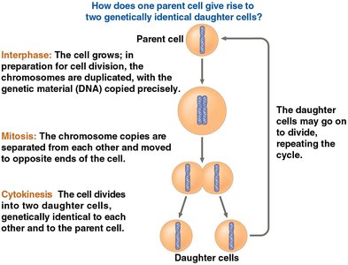
The cell cycle is the series of events that take place in a cell leading to its division and duplication. It is fundamental to reproduction, growth, and repair in multicellular organisms. The cell cycle consists of two main phases: Interphase (cell growth and DNA replication) and the Mitotic (M) phase (nuclear and cytoplasmic division).
Reproduction: Cell division enables organisms to reproduce, either asexually (unicellular) or as part of development (multicellular).
Growth and Development: Multicellular organisms grow by increasing cell number through division.
Repair: Damaged tissues are repaired by cell division.
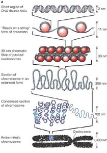
Genetic Material Organization
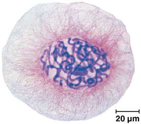
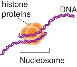
The genome is all the DNA in a cell. In prokaryotes, the genome is a single DNA molecule; in eukaryotes, it consists of multiple DNA molecules organized into chromosomes. Chromosomes are made of DNA and proteins (mainly histones), forming a complex called chromatin. During cell division, chromatin condenses to form visible chromosomes.
Somatic cells: Non-reproductive cells with two sets of chromosomes.
Gametes: Reproductive cells (sperm and eggs) with one set of chromosomes.
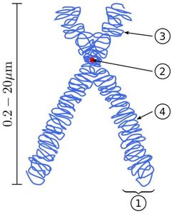
Chromatid: One of two identical halves of a duplicated chromosome.
Phases of the Cell Cycle
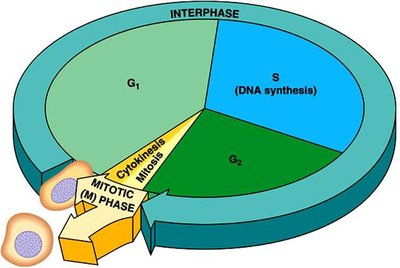
The cell cycle is divided into:
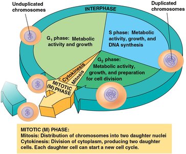
Interphase (about 90% of the cycle):
G1 phase (First Gap): Cell growth and normal metabolic roles.
S phase (Synthesis): DNA replication; chromosomes are duplicated.
G2 phase (Second Gap): Further growth and preparation for mitosis.
Mitotic (M) Phase: Includes mitosis (nuclear division) and cytokinesis (cytoplasmic division).
Mitosis: Division of the Nucleus
Phases of Mitosis
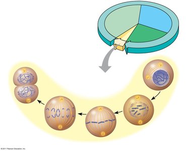
Mitosis is conventionally divided into five sub-phases:
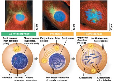
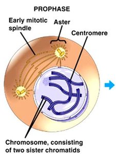
Prophase: Chromatin condenses into chromosomes; mitotic spindle begins to form; nucleolus disappears.
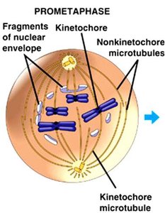
Prometaphase: Nuclear envelope fragments; spindle microtubules attach to kinetochores on chromosomes.
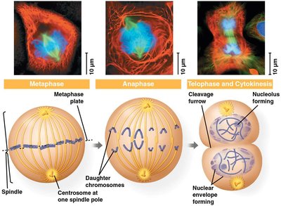
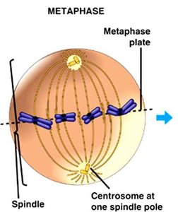
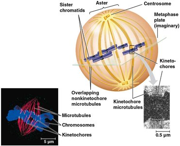
Metaphase: Chromosomes align at the metaphase plate; spindle is fully formed.
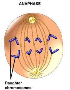
Anaphase: Sister chromatids separate and move toward opposite poles.
Telophase: Nuclear envelopes reform; chromosomes decondense; cytokinesis begins.
Key Structures in Mitosis
Centrosome: Microtubule organizing center; duplicates during interphase.
Mitotic spindle: Structure made of microtubules that segregates chromosomes.
Aster: Radial array of short microtubules from each centrosome.
Kinetochore: Protein complex at the centromere where spindle fibers attach.
Centromere: Region linking sister chromatids.
Detailed Events of Mitosis
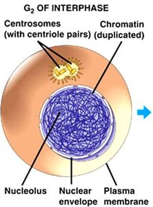
G2 of Interphase
Nuclear envelope and nucleolus are intact.
DNA is duplicated but not yet condensed.
Centrosomes are duplicated.
Prophase
Chromatin condenses into visible chromosomes.
Mitotic spindle begins to form.
Nucleolus disappears.
Prometaphase
Nuclear envelope fragments.
Spindle microtubules attach to kinetochores.
Metaphase
Chromosomes align at the metaphase plate.
Each chromatid is attached to spindle fibers from opposite poles.
Anaphase
Sister chromatids separate and move to opposite poles.
Cell elongates due to non-kinetochore microtubules.
Telophase and Cytokinesis
Nuclear envelopes reform around chromosomes.
Chromosomes decondense.
Cytokinesis divides the cytoplasm, forming two daughter cells.
Regulation of the Cell Cycle
Cell Cycle Control System
The cell cycle is regulated by a control system with checkpoints at critical points (G1, G2, and M phases). Progression through the cycle depends on receiving "go-ahead" signals at these checkpoints.
G1 checkpoint: Determines if the cell will divide.
G2 checkpoint: Checks for DNA damage and completion of replication.
M checkpoint: Ensures all chromosomes are attached to the spindle before anaphase.
Regulatory Molecules
Cyclins: Proteins whose concentrations fluctuate during the cell cycle.
Cyclin-dependent kinases (Cdks): Enzymes that are active only when bound to cyclins; regulate cell cycle events by phosphorylating target proteins.
MPF (Maturation-Promoting Factor): A cyclin-Cdk complex that triggers the cell's passage from G2 to M phase.
Internal and External Signals
Internal signals: Include the presence of enzymes, completion of DNA replication, and proper spindle attachment.
External signals: Include growth factors (e.g., PDGF), nutrient availability, density-dependent inhibition, and anchorage dependence.
Cancer and Loss of Cell Cycle Control
Cancer cells: Do not respond to normal cell cycle controls; divide uncontrollably.
Transformation: Process by which a normal cell becomes cancerous.
Tumor: Mass of abnormal cells; can be benign (localized) or malignant (invasive, metastatic).
Summary: The cell cycle is a tightly regulated process essential for growth, development, and tissue repair. Its regulation ensures genetic stability, and its disruption can lead to diseases such as cancer.
