 Back
BackThe Evolution of Microbial Life: Viruses, Prokaryotes, and Protists
Study Guide - Smart Notes
 Tailored notes based on your materials, expanded with key definitions, examples, and context.
Virus Structure and Replication
Virus Structure
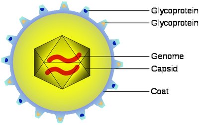
Viruses are acellular infectious agents much smaller than cells, consisting of genetic material encased in a protein shell. They rely on host cells for replication and metabolism.
Capsid: The protein coat that surrounds the viral genome, composed of subunits called capsomeres.
Genetic material can be double- or single-stranded DNA or RNA.
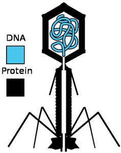
Bacteriophages: Viruses that infect bacteria, often with complex capsids.
Viral envelope: A lipid membrane derived from the host cell, common in animal viruses.
Host range: The spectrum of host cells a virus can infect, determined by specific interactions between viral surface proteins and host cell receptors.
Virus Replication
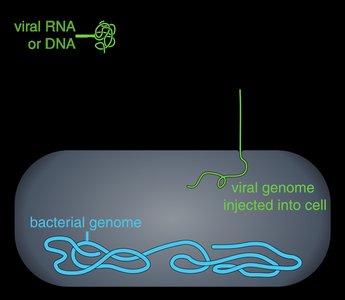
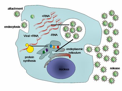
Viral replication begins with attachment to a host cell, followed by entry of the viral genome. The host cell's machinery is hijacked to produce viral components, which self-assemble into new viruses.
Entry mechanisms include direct injection (bacteriophages), endocytosis, or membrane fusion (animal viruses).
The host provides nucleotides, enzymes, ribosomes, tRNA, amino acids, and ATP for viral replication.
New viral particles are assembled and released from the host cell.
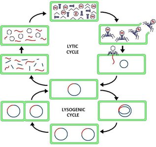
Lytic and Lysogenic Cycles
Bacteriophages can replicate via two main cycles:
Lytic cycle: The phage replicates and lyses (kills) the host cell, releasing new phages. Virulent phages use only this cycle.
Lysogenic cycle: The phage genome integrates into the host chromosome as a prophage and is replicated along with the host without killing it. Temperate phages can switch between cycles.
Bacteria may use restriction enzymes to degrade viral DNA as a defense.
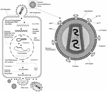
Animal Virus Replication and Retroviruses
Animal viruses often have envelopes and RNA genomes. Replication involves entry, synthesis of viral proteins and genomes, and assembly.
Retroviruses: RNA viruses that use reverse transcriptase to synthesize DNA from their RNA genome, integrating into the host genome as a provirus.
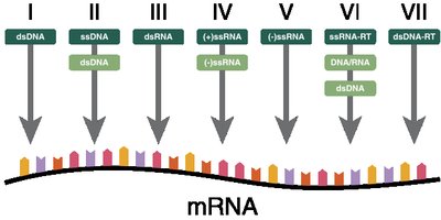
Types of Viral Genomes
Double-stranded DNA viruses: Replicate in the nucleus, often during S phase.
Double-stranded RNA viruses: Replicate in the cytosol using viral enzymes.
Positive-sense RNA viruses: Genome acts as mRNA for immediate translation.
Negative-sense RNA viruses: Genome is complementary to mRNA; requires viral RNA polymerase for transcription.
Retroviruses: (+)ssRNA viruses that reverse transcribe their genome into DNA.
Other Infectious Agents: Viroids and Prions
Viroids: Small, circular, single-stranded RNA molecules that infect plants, do not encode proteins, and replicate using host enzymes.
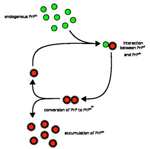
Prions: Infectious proteins that cause neurodegenerative diseases by inducing misfolding of normal proteins.
Prokaryotic Cell Structure and Diversity
Prokaryotic vs. Eukaryotic Cells
Cells are classified as prokaryotic or eukaryotic based on the presence of a nucleus and membrane-bound organelles.
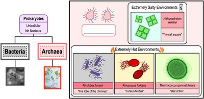
Prokaryotic cells: Lack a nucleus and membrane-bound organelles; include Bacteria and Archaea.
Eukaryotic cells: Have a nucleus and organelles; include plants, animals, fungi, and protists.
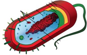
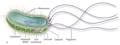
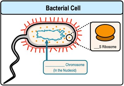
Features of Bacterial Cells
Bacteria are the most abundant and diverse organisms on Earth.
Bacterial DNA is circular and located in the nucleoid region.
Divide by binary fission and have small (70S) ribosomes.
Archaea
Archaea are prokaryotes with unique rRNA sequences and cell walls lacking peptidoglycan.
Many are extremophiles, thriving in extreme environments (high salt, temperature, acidity).
Bacterial Cell Wall Structure
Cell wall: Semi-rigid structure outside the cell membrane, provides protection from osmotic pressure.
Peptidoglycan: Main component of bacterial cell walls, provides rigidity.
Bacteria are classified as Gram-positive (thick peptidoglycan) or Gram-negative (thin peptidoglycan, outer membrane) based on Gram staining.
Glycocalyx: Capsules and Slime Layers
Glycocalyx: Gel-like polysaccharide layer outside the cell wall, aids in attachment and protection from dehydration.
Capsule: Organized, tightly attached layer.
Slime layer: Unorganized, loosely attached layer.
Important for biofilm formation.
Pili and Fimbriae
Pili: Short, filamentous protein structures for attachment, motility, and DNA transfer (conjugation).
Fimbriae: Shorter and more numerous than pili, involved in adhesion and biofilm formation.
Endospores
Endospores: Dormant, highly resistant cells formed by some Gram-positive bacteria (e.g., Bacillus, Clostridium) for survival under harsh conditions.
Not a form of reproduction, but a survival mechanism.
Prokaryotic Motility: Flagella and Chemotaxis
Flagella: Long, whip-like structures for motility, composed of flagellin.
Chemotaxis: Movement toward or away from chemical stimuli; positive (toward attractant) or negative (away from repellent).
Phototaxis: Movement in response to light.
Prokaryotic Reproduction and Genetic Exchange
Binary Fission
Prokaryotes reproduce asexually by binary fission, producing two genetically identical daughter cells.
Plasmids
Plasmids: Small, circular DNA molecules that replicate independently of the chromosome, often carry genes for antibiotic resistance.
Can be lost by plasmid curing.
Horizontal Gene Transfer
Genetic exchange between organisms not in a parent-offspring relationship, increasing genetic diversity.
Three main mechanisms:
Transformation: Uptake of free DNA from the environment by competent cells.
Transduction: Transfer of DNA via bacteriophage (virus).
Conjugation: Direct transfer of DNA between cells via a sex pilus, often involving conjugative plasmids (e.g., F plasmid in E. coli).
Hfr Conjugation
Hfr (High Frequency of Recombination) cells have the F plasmid integrated into their chromosome, allowing transfer of chromosomal genes during conjugation.
Prokaryotic Metabolism and Ecology
Nutritional Classification
Organisms are classified by energy, electron, and carbon sources:
Phototrophs: Use light as energy source.
Chemotrophs: Use chemicals as energy source.
Lithotrophs: Use inorganic molecules as electron source.
Organotrophs: Use organic molecules as electron source.
Autotrophs: Use CO2 as carbon source (carbon fixation).
Heterotrophs: Use organic molecules as carbon source.
Oxygen Requirements
Obligate aerobes: Require O2 for growth.
Anaerobes: Grow without O2.
Facultative anaerobes: Can grow with or without O2.
Aerotolerant anaerobes: Indifferent to O2.
Microaerophiles: Require low O2 concentrations.
Biofilms
Biofilms: Communities of microbes encased in a polysaccharide matrix (extracellular polymeric substances, EPS) attached to surfaces.
Biofilms can include bacteria, archaea, and other microbes, and are medically significant due to their resistance to antibiotics.
Prokaryotic Diversity and Environmental Adaptations
Major Prokaryotic Lineages
Proteobacteria: Diverse group of Gram-negative bacteria, includes nitrogen fixers and pathogens.
Chlamydiae: Gram-negative, lack peptidoglycan, obligate intracellular parasites.
Spirochetes: Gram-negative, spiral-shaped heterotrophs, some are pathogenic.
Cyanobacteria: Gram-negative photoautotrophs, only bacteria to perform oxygenic photosynthesis.
Actinobacteria: High-GC Gram-positive bacteria, includes antibiotic-producing Streptomyces.
Firmicutes: Low-GC Gram-positive bacteria, includes Lactobacillus.
Salt Tolerance
Non-halotolerant: Cannot tolerate moderate salt concentrations.
Halotolerant: Can tolerate moderate salt concentrations.
Halophiles: Require moderate to high salt concentrations (1-14%).
Extreme halophiles: Require very high salt concentrations (>15%).
Environmental Classifications
Microbes are also classified by temperature (e.g., mesophiles, thermophiles, hyperthermophiles), pH (acidophiles, alkaliphiles), and oxygen requirements.
Prokaryotes in the Environment
Ecological Roles
Prokaryotes are essential for nutrient cycling (e.g., nitrogen fixation, oxygen production by cyanobacteria).
They also play roles in human health (microbiome) and disease (pathogens).
The Human Microbiome
Microbiome: Communities of microbes living on/in humans, including resident and transient microbiota.
Symbiotic relationships can be mutualistic, commensal, or pathogenic.
Pathogenic Toxins and Virulence
Toxins: Biological poisons produced by pathogens, classified as exotoxins (proteins released by bacteria) or endotoxins (LPS from Gram-negative bacteria).
Virulence: Degree of pathogenicity; virulence factors are traits that enhance a pathogen's ability to cause disease (e.g., adhesins, capsules, toxins).
Introduction to Protists
What is a Protist?
Protists are a diverse group of eukaryotic organisms that are not plants, animals, or fungi.
Most are unicellular, but some are multicellular or colonial.
They exhibit a wide range of nutritional strategies (heterotrophic, autotrophic, mixotrophic).
Diversity of Protist Structure and Function
Protists have standard eukaryotic organelles, but may possess unique structures.
Most live in aquatic or moist environments.
Some can switch between heterotrophy and autotrophy (mixotrophy).
Evolution of Protists: Endosymbiosis
Primary endosymbiosis: Eukaryotes originated when a host cell engulfed a prokaryote (e.g., origin of mitochondria and chloroplasts).
Secondary endosymbiosis: Eukaryotic cells engulfed other eukaryotic cells, leading to complex plastids with multiple membranes.
Protist Life Cycles
Protists exhibit diverse life cycles, including sexual and asexual reproduction, alternation of generations, and complex host requirements (e.g., Plasmodium in malaria).
Examples: Alternation of generations in Laminaria, conjugation and binary fission in Paramecium.
Eukaryotic Supergroups
Modern classification groups eukaryotes into four supergroups: Excavata, SAR, Archaeplastida, and Unikonta, based on genetic and morphological evidence.
