 Back
The Nervous System: Structure, Function, and Disorders
Study Guide - Smart Notes
 Tailored notes based on your materials, expanded with key definitions, examples, and context.
Nervous System Overview
Characteristics of the Nervous System
The nervous system is a highly specialized network responsible for receiving, integrating, and responding to information from both internal and external environments. It works closely with the endocrine system to maintain homeostasis and is essential for self-awareness and emotional responses.
Receives information from multiple sources simultaneously.
Integrates information to process and make sense of stimuli.
Responds rapidly, often within tenths of a second.
Initiates specific responses such as muscle contraction, glandular secretion, and conscious movement.
Enables self-awareness and emotions.
Organization of the Nervous System
Principal Divisions
The nervous system is divided into two main parts: the central nervous system (CNS) and the peripheral nervous system (PNS).
Central Nervous System (CNS): Consists of the brain and spinal cord. Responsible for receiving, processing, and transferring information.
Peripheral Nervous System (PNS): Composed of nerves outside the CNS. Subdivided into:
Sensory division: Carries information toward the CNS.
Motor division: Carries information away from the CNS.
Somatic division: Controls skeletal muscles (voluntary).
Autonomic division: Controls smooth muscle, cardiac muscle, and glands (involuntary). Includes sympathetic and parasympathetic divisions.
Neurons: The Communication Cells
Structure and Function of Neurons
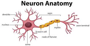
Neurons are specialized cells that generate and conduct electrical impulses, forming the basis of nervous system communication. Each neuron consists of three main parts:
Cell body (soma): Contains the nucleus and most organelles.
Dendrites: Receive incoming information from other neurons or sensory receptors.
Axon: Conducts electrical impulses away from the cell body toward target cells.
Sensory neurons: Transmit information from sensory receptors to the CNS.
Interneurons: Transmit information within the CNS.
Motor neurons: Transmit information from the CNS to effectors (muscles or glands).
Action Potentials
Neurons communicate via action potentials, which are rapid electrical impulses that travel along the axon. Action potentials are the primary means of communication in the nervous system.
All-or-none principle: An action potential occurs only if a threshold is reached; it is always the same speed and voltage.
Self-propagating: Once initiated, the action potential moves down the axon without decreasing in strength.
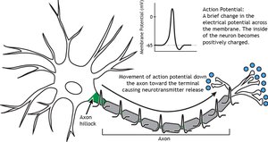
Equation: The change in membrane potential during an action potential can be described by the Nernst equation:
Neuroglial Cells
Neuroglial (glial) cells support and protect neurons but do not transmit action potentials. They make up about 80% of nervous system cells.
Schwann cells: A type of glial cell in the PNS that forms the myelin sheath around axons, aiding in faster impulse transmission.
Synapses and Neurotransmitters
Synaptic Transmission
A synapse is a specialized junction where an axon terminal communicates with its target cell (another neuron, muscle cell, or gland). Neurotransmitters are chemical messengers released into the synaptic cleft to transmit signals.
Excitatory neurotransmitters: Depolarize the postsynaptic cell, increasing the likelihood of an action potential.
Inhibitory neurotransmitters: Hyperpolarize the postsynaptic cell, decreasing the likelihood of an action potential.
Neurotransmitter removal: Occurs by reuptake, enzymatic destruction, or diffusion away from the synaptic cleft.
Divisions of the Peripheral Nervous System
Somatic Motor Division
The somatic motor division controls voluntary movements of skeletal muscles and mediates involuntary reflexes.
Voluntary control: Conscious movement of skeletal muscles.
Reflexes: Involuntary responses, such as the stretch reflex (e.g., knee-jerk reflex) and crossed extensor reflex.
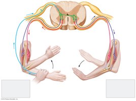
Example: The patellar (knee-jerk) reflex is a classic stretch reflex used by physicians to assess nervous system function.
Autonomic Division
The autonomic division regulates involuntary functions of internal organs and consists of two antagonistic branches:
Sympathetic division: Prepares the body for emergencies (fight-or-flight response). Key neurotransmitter: norepinephrine.
Parasympathetic division: Promotes relaxation and maintenance activities (rest-and-digest). Key neurotransmitter: acetylcholine.
Both divisions innervate smooth muscle, cardiac muscle, and glands, often producing opposite effects to maintain homeostasis.
Central Nervous System: Brain and Spinal Cord
Protection of the CNS
The CNS is protected by several structures:
Bone: Skull and vertebrae encase the brain and spinal cord.
Meninges: Three protective membranes (dura mater, arachnoid, pia mater).
Cerebrospinal fluid: Cushions the brain and spinal cord, acts as a shock absorber, and is produced in the brain's ventricles.
Blood-brain barrier: Restricts entry of certain chemicals and pathogens into the CNS.
Spinal Cord Structure and Function
The spinal cord serves as a major pathway for transmitting information between the brain and the rest of the body. It is protected by vertebrae and surrounded by meninges and cerebrospinal fluid.
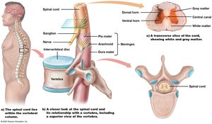
Relays sensory and motor information to and from the brain.
Coordinates reflexes independent of the brain.
Brain Structure and Function
The brain is the command center of the body, divided into three major regions:
Hindbrain: Includes the medulla oblongata (controls vital functions), cerebellum (coordinates movement), and pons (relays information).
Midbrain: Coordinates head movements, vision, hearing, and sleep/wake cycles.
Forebrain: Involved in emotions, conscious thought, and higher cognitive functions. Includes the hypothalamus (homeostasis), thalamus (sensory relay), limbic system (emotions, memory), and cerebrum (language, decision making).
Cerebral cortex: The outer layer of the cerebrum, responsible for conscious thought, sensory perception, voluntary movement, memory, and understanding.
Disorders of the Nervous System
Types of Disorders
Trauma: Includes concussions and spinal cord injuries, which can impair sensation and function.
Infections: Such as encephalitis (brain inflammation) and meningitis (meningeal inflammation), caused by viruses or bacteria.
Neural and synaptic transmission disorders: Examples include epilepsy, Alzheimer's disease, and Parkinson's disease.
Concussion
A concussion is a type of traumatic brain injury resulting from a blow to the head. It can cause headaches, memory problems, and coordination issues. Severe cases may lead to subdural hematoma, requiring surgical intervention.
Spinal Cord Injuries
Injuries to the spinal cord can result in loss of sensation and function below the site of injury. The severity depends on the location of the injury:
Paraplegia: Paralysis of the legs and lower trunk (injuries below the neck).
Quadriplegia: Paralysis of all four limbs and trunk (injuries to the neck).
Encephalitis and Meningitis
Encephalitis: Inflammation of the brain, often viral (e.g., herpes simplex, West Nile virus). Symptoms range from mild to severe and may include fever, confusion, and seizures. Treated with antiviral drugs.
Meningitis: Inflammation of the meninges, caused by bacteria or viruses. Bacterial meningitis is life-threatening and requires antibiotics; viral meningitis is usually milder.
Alzheimer’s Disease
Alzheimer’s disease is the most common cause of dementia, characterized by progressive memory loss and personality changes. It is associated with abnormal protein accumulation (tau and beta amyloid) in the brain, affecting the hippocampus and cerebral cortex.
Diagnosis: Based on symptoms; research is ongoing for better diagnostic tests and treatments.
