 Back
The Urinary System: Structure, Function, and Homeostasis
Study Guide - Smart Notes
 Tailored notes based on your materials, expanded with key definitions, examples, and context.
The Urinary System: Overview and Functions
Introduction to the Urinary System
The urinary system is essential for maintaining homeostasis by regulating the composition, volume, and pressure of blood. Its primary organs are the kidneys, which filter blood, remove metabolic wastes, and balance water and electrolytes. The system also plays a role in hormone secretion and acid-base balance.
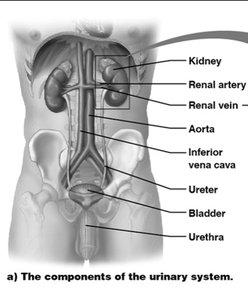
Primary Organs: Kidneys, ureters, bladder, and urethra.
Main Functions: Excretion of wastes, regulation of fluid and electrolyte balance, maintenance of blood pH, and hormone secretion (erythropoietin and renin).
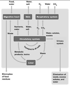
Functional Relationships of the Urinary System
Homeostasis and Fluid Balance
Physiological systems operate in a fluid environment, requiring the regulation of water and solute concentrations. The urinary system, through osmoregulation, maintains these concentrations within narrow limits, balancing water gain and loss.
Osmoregulation: The process of controlling solute concentrations and water balance.
Homeostasis: The urinary system works with other organ systems to maintain internal stability.
Excretory Processes and Nitrogenous Waste
Key Functions of Excretory Systems
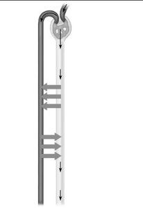
Most excretory systems, including the human urinary system, produce urine by refining a filtrate derived from body fluids. The main processes are filtration, reabsorption, secretion, and excretion.
Filtration: Filtering of body fluids to form a filtrate.
Reabsorption: Reclaiming valuable solutes from the filtrate back into the blood.
Secretion: Adding nonessential solutes and wastes to the filtrate.
Excretion: Removal of processed filtrate (urine) from the body.
Nitrogenous Wastes
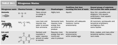
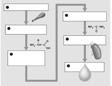
Excretory systems eliminate nitrogenous wastes resulting from protein digestion. These wastes are excreted as ammonia, urea, or uric acid, depending on the organism and its environment.
Nitrogenous waste | Chemical formula | Advantages | Disadvantages | Conditions favoring excretion | Organisms |
|---|---|---|---|---|---|
Ammonia | NH3 | Takes almost no energy to produce | Highly toxic | Aquatic | Bony fish, crocodiles, aquatic amphibians |
Urea | (NH2)2CO | Less toxic, requires less water than ammonia | Somewhat toxic, requires energy to produce | Terrestrial, with adequate water | Mammals, amphibians, some reptiles |
Uric acid | C5H4N4O3 | Nontoxic, insoluble in water, minimal water loss | Requires more energy to produce | Dry terrestrial environments | Birds, reptiles, insects |
Conversion of Ammonia to Urea
In mammals, ammonia produced from amino acid breakdown is converted to urea in the liver, which is less toxic and can be safely transported in the blood to the kidneys for excretion.
Ammonia (NH3): Highly toxic, converted to urea in the liver.
Urea: Less toxic, excreted in urine.
Structural Components of the Urinary System
Gross Anatomy of the Kidneys
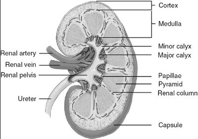
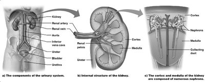
The kidneys are retroperitoneal organs located against the posterior abdominal wall. Each kidney is supplied by a renal artery and drained by a renal vein. The ureter carries urine from the kidney to the bladder.
Major Structures: Cortex, medulla, pyramids, renal columns, papillae, calyces, pelvis, capsule.
Microscopic Anatomy: The Nephron
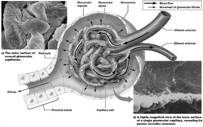
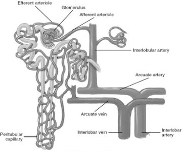
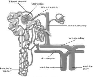
The nephron is the functional unit of the kidney, responsible for urine formation. Each nephron consists of a renal corpuscle (glomerulus and glomerular capsule) and a renal tubule (proximal convoluted tubule, nephron loop, distal convoluted tubule).
Renal Corpuscle: Site of filtration; includes the glomerulus and Bowman’s capsule.
Renal Tubule: Site of reabsorption and secretion; includes proximal tubule, loop of Henle, distal tubule, and collecting duct.
Urine Formation: Processes in the Nephron
Glomerular Filtration
Filtration occurs in the renal corpuscle, where blood pressure forces water and small solutes from the glomerulus into the Bowman’s capsule, forming the filtrate. Large molecules and blood cells are retained in the blood.
Driving Force: Blood pressure.
Filtrate: Contains water, ions, glucose, amino acids, and wastes.
Tubular Reabsorption
Reabsorption moves substances from the filtrate back into the blood, primarily in the proximal tubule. Most nutrients, ions, and water are reabsorbed by active transport and osmosis.
Key Substances Reabsorbed: Salts, glucose, amino acids, water.
Water Reabsorption: Occurs by osmosis except in the ascending limb of the nephron loop, which is impermeable to water.
Tubular Secretion
Secretion is the active transport of substances from the blood into the filtrate. This process removes excess ions (e.g., H+, K+), metabolic wastes, and drugs from the body.
Examples of Secreted Substances: Hydrogen ions, potassium ions, creatinine, histamine, penicillin.
Regulation of Kidney Function and Homeostasis
Hormonal Regulation
The kidneys regulate blood volume and pressure through the secretion of hormones and enzymes:
Renin: Initiates the renin-angiotensin-aldosterone system, increasing blood pressure and volume.
Aldosterone: Increases sodium and water reabsorption.
Antidiuretic Hormone (ADH): Increases water reabsorption in the distal tubule and collecting duct.
Atrial Natriuretic Hormone: Promotes excretion of sodium and water, lowering blood pressure.
Characteristics of Urine
Normal and Abnormal Constituents
Fresh urine is about 95% water and 5% solutes. Its color, clarity, and composition can provide important health information.
Normal Color: Clear yellow, varies with hydration.
Specific Gravity: 1.001 to 1.035.
pH: 4.6 to 8.0.
Abnormal Constituents: Albumin, glucose, blood cells, ketone bodies, drugs, microbes.
Urine Color and Clinical Significance
Color | Possible Cause | Necessary Action |
|---|---|---|
Yellow/Gold | Normal, hydration status, B vitamins | None |
Red/Pink | Hematuria, foods, drugs | Consult physician if blood suspected |
White/Colorless | Excess hydration | Consult if chronic |
Orange | Dehydration, foods, drugs, liver issues | Drink water, consult if persists |
Amber/Brown | Severe dehydration, liver/kidney issues | Consult physician |
Green/Blue | Rare, drugs, foods, UTIs | Consult if persistent or symptomatic |
Cloudy/Sediment/Foamy | UTI, proteinuria, kidney stones | Consult physician |
Acid-Base and Electrolyte Balance
Blood pH Regulation
The kidneys help maintain blood pH (7.35–7.45) by excreting hydrogen ions and reabsorbing bicarbonate. Acid-base imbalances can be respiratory or metabolic in origin.
Respiratory Acidosis: CO2 retention, decreased pH.
Respiratory Alkalosis: CO2 loss, increased pH.
Metabolic Acidosis: Increased metabolic acids or decreased bicarbonate.
Metabolic Alkalosis: Loss of acids or increased bicarbonate.
Renal Failure and Dialysis
Dialysis as Kidney Replacement Therapy
Dialysis is a medical procedure that substitutes for the filtration function of healthy kidneys in patients with renal failure. There are two main types:
Continuous Ambulatory Peritoneal Dialysis (CAPD): Uses the peritoneum as a filter; can be done at home but carries infection risk.
Hemodialysis: Blood is filtered through a machine at a dialysis center; wastes and excess ions diffuse into dialysis fluid.
Additional info: The urinary system interacts with other organ systems (e.g., cardiovascular, endocrine, respiratory) to maintain overall body homeostasis, as illustrated in the provided diagrams.
