 Back
BackTissues, Organs, and Organ Systems: Structure and Function in Multicellular Organisms
Study Guide - Smart Notes
 Tailored notes based on your materials, expanded with key definitions, examples, and context.
Tissues, Organs, and Organ Systems
Introduction to Biological Organization
Multicellular organisms exhibit a hierarchical organization, where cells are grouped into tissues, tissues form organs, organs combine into organ systems, and organ systems function together to sustain the organism. This organization allows for specialization and division of labor, essential for complex life forms.
Cell: The basic structural and functional unit of life.
Tissue: Groups of similar cells performing a common function.
Organ: Structures composed of multiple tissue types working together.
Organ System: Groups of organs that carry out major body functions.
Organism: An individual living entity composed of multiple organ systems.
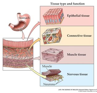
The Four Basic Tissue Types
All organs in the body are constructed from four fundamental tissue types, each with distinct structures and functions:
Epithelial tissue: Covers body surfaces, lines cavities, and forms glands.
Connective tissue: Supports, binds, and protects other tissues and organs.
Muscle tissue: Specialized for contraction and movement.
Nervous tissue: Specialized for communication via electrical and chemical signals.
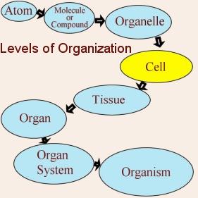
Levels of Organization in Living Beings
The complexity of living organisms arises from the integration of various levels of biological organization. Each level builds upon the previous, allowing for increased specialization and function.
Atoms & Molecules: Chemical building blocks of cells.
Organelles: Specialized structures within cells.
Cells: Basic units of structure and function.
Tissues: Groups of similar cells.
Organs: Composed of multiple tissue types.
Organ Systems: Groups of organs with related functions.
Organism: The complete living entity.
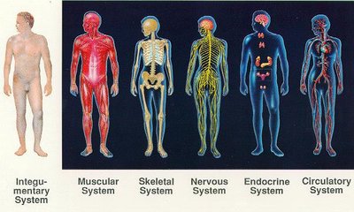
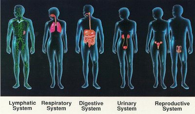
Homeostasis and Organ Systems
Organ systems work together to maintain homeostasis, the stable internal environment necessary for survival. Changes in the external environment can trigger physiological responses to restore balance.
Examples of organ systems: Integumentary, muscular, skeletal, nervous, endocrine, circulatory, lymphatic, respiratory, digestive, urinary, reproductive.
Coordination: Nervous and endocrine systems play key roles in regulating and integrating the activities of other systems.
Epithelial Tissue
Structure and Function
Epithelial tissue consists of closely packed cells with minimal extracellular matrix, forming protective barriers on body surfaces and lining internal cavities. It also forms glands for secretion.
Functions: Protection, absorption, secretion, sensation, and filtration.
Polarity: Epithelial cells have an apical (free) surface and a basal surface attached to a basement membrane.
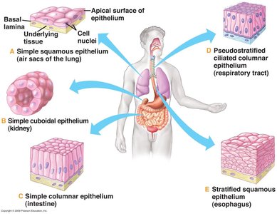
Types of Epithelial Tissue
Simple squamous: Single layer of flat cells (e.g., air sacs of lungs).
Simple cuboidal: Single layer of cube-shaped cells (e.g., kidney tubules).
Simple columnar: Single layer of tall, column-like cells (e.g., intestine).
Pseudostratified ciliated columnar: Appears layered but all cells touch the basement membrane (e.g., respiratory tract).
Stratified squamous: Multiple layers, surface cells flattened (e.g., esophagus).
Specializations of Epithelial Cells
Microvilli: Increase surface area for absorption (e.g., small intestine).
Cilia: Move substances along the surface (e.g., respiratory tract).
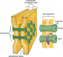
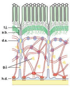
Cell junctions: Tight junctions, desmosomes, gap junctions, and hemidesmosomes provide adhesion and communication between cells.
Glandular Epithelium
Some epithelial cells are specialized to form glands, which synthesize and secrete substances.
Exocrine glands: Secrete products onto body surfaces or into body cavities (e.g., sweat, salivary glands).
Endocrine glands: Secrete hormones directly into the bloodstream (e.g., thyroid, pituitary glands).
Connective Tissue
Structure and Function
Connective tissue supports, binds, and protects other tissues and organs. It is characterized by abundant extracellular matrix (ECM) composed of fibers and ground substance.
Cells: Fibroblasts, adipocytes, chondrocytes, osteocytes, blood cells.
Fibers: Collagen (strength), elastic (flexibility), reticular (support).
Ground substance: Gel-like material containing glycosaminoglycans (GAGs) and proteoglycans.
Types of Connective Tissue
Connective tissue proper: Loose and dense connective tissues.
Specialized connective tissues: Cartilage, bone, blood, adipose tissue.
Adipose Tissue
White adipose tissue: Stores energy, insulates, cushions organs.
Brown adipose tissue: Generates heat, especially in infants and hibernating animals.
Cartilage and Bone
Cartilage: Flexible, avascular tissue with chondrocytes in ECM; types include hyaline, elastic, and fibrocartilage.
Bone: Rigid, mineralized tissue with osteocytes; supports and protects the body, stores minerals.
Blood
Blood is a connective tissue with a liquid ECM (plasma) and various cell types (erythrocytes, leukocytes, platelets). It transports gases, nutrients, wastes, and immune cells.
Muscle Tissue
Structure and Function
Muscle tissue is specialized for contraction, enabling movement of the body and its parts. It contains contractile proteins (actin and myosin) and requires ATP for function.
Skeletal muscle: Voluntary, striated, multinucleated fibers; moves bones.
Cardiac muscle: Involuntary, striated, branched cells; found in the heart.
Smooth muscle: Involuntary, non-striated; found in walls of internal organs and blood vessels.
Nervous Tissue
Structure and Function
Nervous tissue is specialized for rapid communication and control. It consists of neurons (nerve cells) and glial cells (supporting cells).
Neurons: Generate and transmit electrical impulses (action potentials).
Glial cells: Support, insulate, and protect neurons; form myelin sheaths.
Neurons have a cell body, dendrites (receive signals), and an axon (transmits signals). Synapses are specialized junctions for communication between neurons or between neurons and other cells.
Summary Table: The Four Basic Tissue Types
Tissue Type | Main Function | Examples |
|---|---|---|
Epithelial | Protection, absorption, secretion, sensation | Skin, lining of gut, glands |
Connective | Support, binding, protection, transport | Tendons, bone, blood, adipose tissue |
Muscle | Movement, posture, heat production | Skeletal muscles, heart, walls of organs |
Nervous | Communication, control, coordination | Brain, spinal cord, nerves |
