 Back
BackUnit 2: Cells – Structure, Function, Membranes, and Communication
Study Guide - Smart Notes
 Tailored notes based on your materials, expanded with key definitions, examples, and context.
Cells: Structure and Function
Introduction to Cells
Cells are the fundamental units of life, forming the basis of all living organisms. Their structure and function are intricately linked, and understanding these relationships is essential for studying biology at the molecular, cellular, and organismal levels.
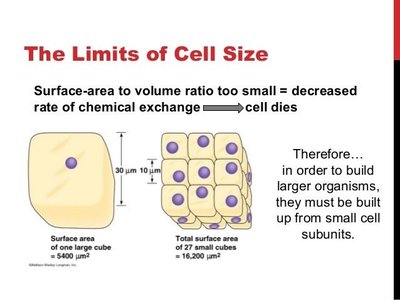
Limits to Cell Size
The size of a cell is constrained by the surface area to volume (SA/V) ratio. As a cell grows, its volume increases faster than its surface area, which limits the efficiency of material exchange with the environment. This principle explains why most cells are small and why larger organisms are composed of many small cells rather than a few large ones.
Surface Area to Volume Ratio: As cell size increases, the SA/V ratio decreases, reducing the rate of chemical exchange and potentially leading to cell death if the cell becomes too large.
Implications: Larger organisms are built from many small cell subunits to maintain efficient exchange with their environment.
Example: Agar cube diffusion experiments demonstrate how diffusion time increases with cell size due to the SA/V ratio.
Biological Adaptations to SA/V Ratio
Organisms have evolved various adaptations to optimize their SA/V ratio for heat exchange and material transport.
Example 1: Human fingers get cold quickly due to a high SA/V ratio, which facilitates rapid heat loss.
Example 2: Elephants have large, thin ears to increase surface area for heat dissipation.


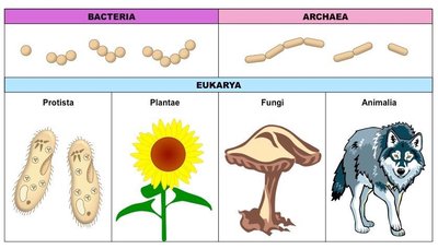
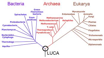
The Three Domains of Life
Classification of Life
All living organisms are classified into three domains: Bacteria, Archaea, and Eukarya. This classification reflects evolutionary relationships and fundamental differences in cell structure.
Bacteria: Prokaryotic cells lacking a nucleus and membrane-bound organelles.
Archaea: Prokaryotic cells with unique membrane lipids and genetic machinery, often found in extreme environments.
Eukarya: Eukaryotic cells with a true nucleus and membrane-bound organelles; includes Protists, Plants, Fungi, and Animals.
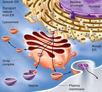
Cell Structure: Organelles and the Endomembrane System
Endomembrane System
The endomembrane system is a network of membranes within eukaryotic cells that work together to modify, package, and transport proteins and lipids.
Key Organelles: Endoplasmic reticulum (smooth and rough), Golgi apparatus, lysosomes, vesicles, and the plasma membrane.
Function: Synthesis, processing, and export of proteins and lipids.
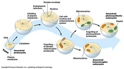
Endosymbiotic Theory
The endosymbiotic theory explains the origin of mitochondria and chloroplasts in eukaryotic cells. According to this theory, these organelles originated as free-living prokaryotes that were engulfed by ancestral eukaryotic cells.
Evidence: Both mitochondria and chloroplasts have their own circular DNA, ribosomes, and double membranes.
Cell Membranes: Structure and Function
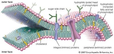
Fluid Mosaic Model
The cell membrane is described by the fluid mosaic model, which depicts the membrane as a dynamic structure composed of a phospholipid bilayer with embedded proteins, cholesterol, and carbohydrates.
Phospholipids: Form the basic structure of the membrane, creating a hydrophobic barrier.
Cholesterol: Modulates membrane fluidity.
Proteins: Facilitate transport, communication, and enzymatic activity.
Carbohydrates: Involved in cell recognition and signaling.
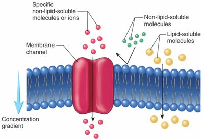
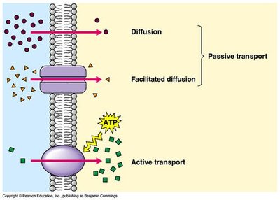
Membrane Transport
Cell membranes are selectively permeable, allowing some substances to cross more easily than others. Transport can be passive or active.
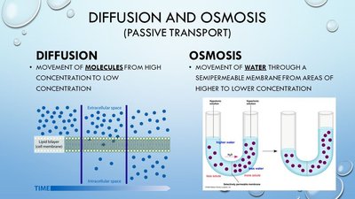
Passive Transport: Movement of molecules down their concentration gradient without energy input (e.g., diffusion, facilitated diffusion, osmosis).
Active Transport: Movement of molecules against their concentration gradient, requiring energy (ATP).
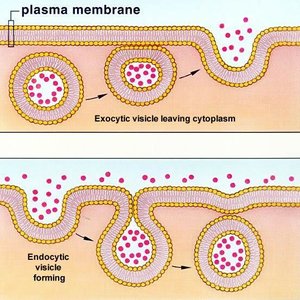
Bulk Transport: Exocytosis and Endocytosis
Large particles or volumes of substances are transported across the membrane via vesicles in processes known as exocytosis (out of the cell) and endocytosis (into the cell).
Exocytosis: Vesicles fuse with the plasma membrane to release contents outside the cell.
Endocytosis: The plasma membrane engulfs material to bring it into the cell.
Osmosis and Water Potential
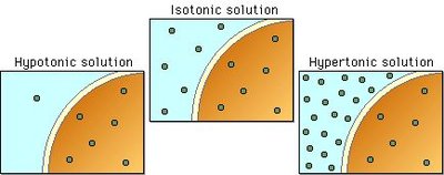
Osmosis and Tonicity
Osmosis is the passive diffusion of water across a semipermeable membrane. Tonicity describes the relative solute concentration of solutions separated by a membrane, affecting water movement.
Hypotonic Solution: Lower solute concentration outside the cell; water enters the cell.
Isotonic Solution: Equal solute concentration; no net water movement.
Hypertonic Solution: Higher solute concentration outside the cell; water leaves the cell.
Water Potential
Water potential (Ψ) predicts the direction of water movement and is determined by solute potential (Ψs) and pressure potential (Ψp):
Formula:
Solute Potential:
Variables: i = ionization constant, C = molarity, R = pressure constant (0.0831 L·bars/mol·K), T = temperature in Kelvin (K = °C + 273)
Pure Water: Has the highest water potential (Ψ = 0); adding solute lowers Ψ.
Example Calculation: Calculate the water potential of a 0.1 M NaCl solution at 25°C in an open container (Ψp = 0):
bars
Cell Communication and Signaling
Overview of Cell Signaling
Cell signaling is essential for the regulation, development, and function of multicellular organisms. It involves the transmission of signals from one cell to another, leading to a specific response.
Stages of Cell Signaling: Reception, Transduction, Response
Reception: A ligand (signal molecule) binds to a receptor.
Transduction: Signal is relayed through a cascade of molecular interactions, often involving kinases and phosphatases (phosphorylation cascades).
Response: Activation or deactivation of cellular processes, such as gene expression or enzyme activity.
The Mitotic Cell Cycle
Phases of the Cell Cycle
The cell cycle is a regulated process that ensures the accurate division of genetic material and cytoplasm, resulting in two genetically identical daughter cells.
Interphase: Includes G1 (cell growth), S (DNA synthesis), and G2 (preparation for mitosis).
Mitosis: Chromosomes condense, align, and are evenly divided.
Cytokinesis: Division of the cytoplasm to form two separate cells.
Regulation of the Cell Cycle
Cell division is tightly regulated by proteins called cyclins and cyclin-dependent kinases (CDKs). Loss of regulation can lead to uncontrolled cell division and cancer.
Cancer: Results from multiple mutations that disrupt normal cell cycle control, leading to benign, malignant, or metastatic tumors.
Summary Table: Key Concepts in Cell Structure and Function
Concept | Definition | Example/Application |
|---|---|---|
Surface Area/Volume Ratio | Ratio of cell membrane area to cell volume | Limits cell size; affects diffusion rates |
Endomembrane System | Network of membranes for protein/lipid processing | ER, Golgi apparatus, vesicles |
Fluid Mosaic Model | Dynamic structure of cell membrane | Phospholipids, proteins, cholesterol |
Osmosis | Diffusion of water across a membrane | Plant cell turgor, red blood cell lysis |
Cell Signaling | Communication between cells via signals | Hormone signaling, immune response |
Cell Cycle | Ordered sequence of cell growth and division | Mitosis, cytokinesis |
