 Back
Circulatory Systems: Structure, Function, and Evolution
Study Guide - Smart Notes
 Tailored notes based on your materials, expanded with key definitions, examples, and context.
CIRCULATORY SYSTEMS
General Functions of Circulatory Systems
The circulatory system is essential for transporting nutrients, gases, and waste products throughout the body. It enables multicellular organisms to maintain homeostasis and supports cellular metabolism.
Transport: Delivers oxygen and nutrients to tissues and removes carbon dioxide and metabolic wastes.
Exchange: Facilitates gas, nutrient, and waste exchange between blood and tissues.
Regulation: Helps regulate temperature, pH, and fluid balance.
Types of Circulatory Systems
Animals possess different types of circulatory systems depending on their complexity and evolutionary lineage.
Gastrovascular Cavity: Found in cnidarians and flatworms; a single opening serves both digestive and circulatory functions.
Open Circulatory System: Seen in arthropods and most molluscs; blood is pumped by a heart into open spaces (sinuses) and directly bathes tissues.
Closed Circulatory System: Found in vertebrates, earthworms, squids, and octopuses; blood is confined to vessels, allowing efficient transport and regulation.
EVOLUTION OF VERTEBRATE CARDIOVASCULAR SYSTEMS
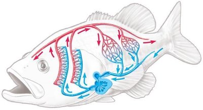
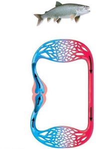
Fish: Single Circulation
Fish possess a two-chambered heart (one atrium, one ventricle) and a single circulatory loop. Blood passes through the heart once per cycle, moving from the heart to the gills for oxygenation and then to the rest of the body.
Single Circulation: Blood flows from heart → gills → body → heart.
Chambers: Atrium receives blood; ventricle pumps blood to gills.
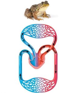
Amphibians: Double Circulation with Three-Chambered Heart
Amphibians have a three-chambered heart (two atria, one ventricle) and double circulation. Blood is pumped twice: once to the lungs/skin (pulmocutaneous circuit) and once to the body (systemic circuit).
Double Circulation: Pulmocutaneous circuit (lungs/skin) and systemic circuit (body).
Mixing: Some mixing of oxygen-rich and oxygen-poor blood occurs in the ventricle.
Reptiles: Partial Division of the Ventricle
Reptiles (except crocodilians) have a three-chambered heart with a partially divided ventricle, reducing mixing of blood. Crocodilians have a four-chambered heart.
Partial Division: Less mixing of oxygen-rich and oxygen-poor blood.
Four Chambers in Crocodilians: Complete separation of circuits.
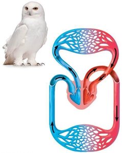
Birds and Mammals: Four-Chambered Heart
Birds and mammals have a four-chambered heart (two atria, two ventricles) with complete separation of pulmonary and systemic circuits. This allows efficient oxygen delivery and supports high metabolic rates.
Right Side: Pumps oxygen-poor blood to lungs (pulmonary circuit).
Left Side: Pumps oxygen-rich blood to body (systemic circuit).
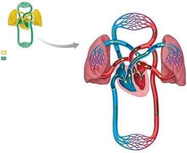
THE HUMAN CARDIOVASCULAR SYSTEM AND HEART
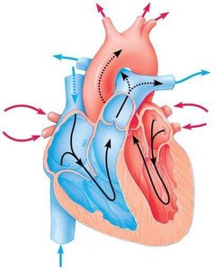
Pathway of Blood Through the Mammalian Heart
Blood flows through the heart in a specific sequence, ensuring oxygenation and distribution to tissues.
Blood enters the right atrium from the body via the superior and inferior vena cava.
Moves to the right ventricle, then pumped to the lungs via the pulmonary artery.
Returns oxygenated to the left atrium via the pulmonary vein.
Moves to the left ventricle, then pumped to the body via the aorta.
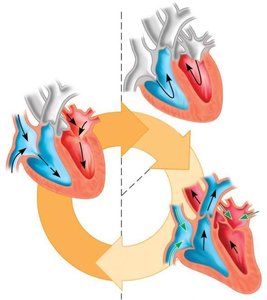
Cardiac Cycle: Diastole and Systole
The heart contracts and relaxes in a rhythmic sequence called the cardiac cycle, consisting of two main phases:
Diastole: Heart is relaxed; blood flows into chambers.
Systole: Heart contracts; blood is pumped out of chambers.
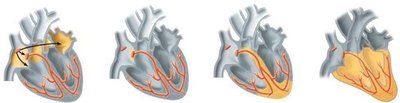
Control of Heartbeats
The heart's rhythm is controlled by specialized cells:
SA Node (Sinoatrial): Pacemaker; initiates electrical impulses.
AV Node (Atrioventricular): Relays signals to ventricles.
Electrocardiogram (ECG): Measures electrical activity of the heart.
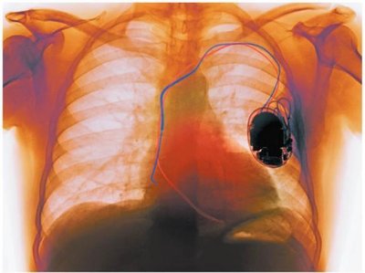
Artificial Pacemaker: Device used when natural pacing fails.
HEART ATTACKS AND CARDIOVASCULAR DISEASE
Heart Attack and Stroke
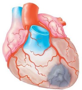
A heart attack is the damage or death of cardiac muscle tissue, usually caused by a blocked coronary artery. A stroke is the death of brain tissue due to lack of oxygen from blocked or ruptured arteries.
Coronary Arteries: Supply heart muscle with oxygen and nutrients.
Blockage: Leads to tissue death and impaired function.
Atherosclerosis
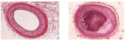
Atherosclerosis is a chronic cardiovascular disease characterized by the buildup of fatty plaques in arteries, narrowing blood flow and increasing risk of heart attack and stroke.
Plaques: Fatty deposits in arterial walls.
Inflammation: Associated with plaque formation and clotting.
STRUCTURE AND FUNCTION OF BLOOD VESSELS
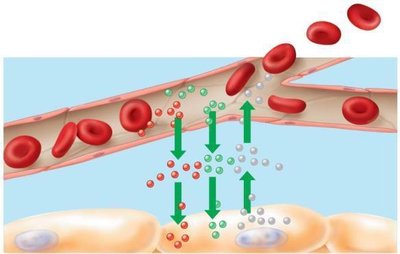
Capillaries
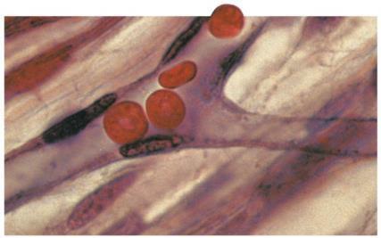
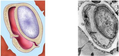
Capillaries are the smallest blood vessels, with thin walls for efficient exchange of gases, nutrients, and wastes between blood and tissues.
Structure: Single layer of epithelial cells; narrow diameter.
Function: Exchange with interstitial fluid.

Arteries and Veins
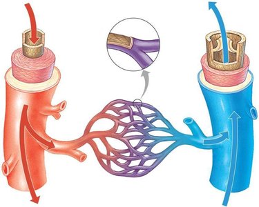
Arteries and veins are larger vessels with distinct structural features:
Arteries: Thick smooth muscle; elastic fibers; carry blood away from heart.
Veins: Thinner walls; valves to prevent backflow; carry blood to heart.
Blood Pressure and Velocity
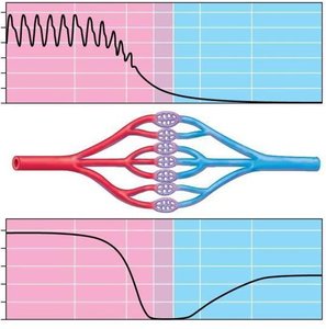
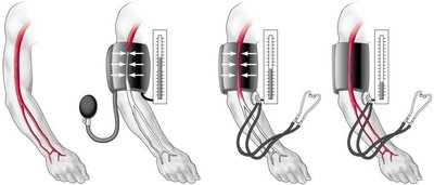
Blood pressure is the force exerted by blood on vessel walls, highest in arteries and lowest in veins. Velocity decreases in capillaries to allow exchange.
Systolic Pressure: Pressure during ventricular contraction.
Diastolic Pressure: Pressure between contractions.
Venous Return
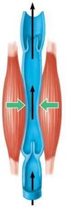
Blood returns to the heart through veins, aided by muscle contractions and one-way valves.
Muscle Contractions: Squeeze veins, pushing blood toward heart.
Valves: Prevent backward flow.
Measuring Blood Pressure
Blood pressure is measured using a sphygmomanometer. Normal adult blood pressure is about 120/70 mm Hg; hypertension is persistent high blood pressure (≥140/90 mm Hg).
Hypertension: Increases risk of heart attack, stroke, and kidney failure.
Regulation of Blood Flow
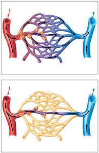
Smooth muscle in arteriole walls and precapillary sphincters regulate blood flow and pressure.
Precapillary Sphincters: Control blood entry into capillary beds.
Capillary Exchange
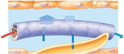
Substances move across capillary walls by diffusion, vesicle transport, and pressure-driven flow. Blood pressure forces fluid out at the arterial end; osmotic pressure draws fluid in at the venous end.
Lymphatic System: Picks up excess fluid from tissues.
STRUCTURE AND FUNCTION OF BLOOD
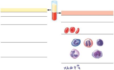
Components of Blood
Blood is composed of plasma and cellular elements, each with specific functions.
Plasma: 90% water; contains ions, proteins, nutrients, wastes, gases, and hormones.
Red Blood Cells (Erythrocytes): Transport oxygen bound to hemoglobin.
White Blood Cells (Leukocytes): Defend against infection; include phagocytes (monocytes, neutrophils).
Platelets: Cell fragments involved in blood clotting.
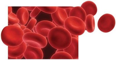
Red Blood Cells: Structure, Function, and Production
Red blood cells are essential for oxygen transport. They are produced in bone marrow and recycled after 3-4 months.
Anemia: Caused by low hemoglobin or red cell count; results in fatigue.
Erythropoietin (EPO): Hormone stimulating red cell production.
Blood Doping: Artificially increasing red cell count; risks include clotting and heart failure.
Blood Clotting
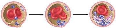
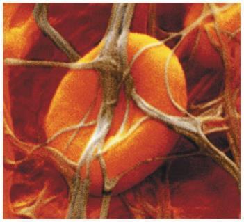
Blood clotting prevents blood loss when vessels are injured. Platelets adhere to exposed tissue, forming a plug, and clotting factors produce fibrin threads to reinforce the clot.
Platelet Plug: Initial response to injury.
Fibrin Clot: Final reinforced patch.
Stem Cells and Blood Cell Diseases
Stem cells in bone marrow differentiate into various blood cells. Leukemia is cancer of white blood cells, treated by radiation, chemotherapy, or bone marrow transplant.
Lymphoid Stem Cells: Produce lymphocytes for immune function.
Myeloid Stem Cells: Produce erythrocytes, other leukocytes, and platelets.
Summary Table: Types of Circulatory Systems
Type | Main Features | Examples |
|---|---|---|
Gastrovascular cavity | Single opening, direct exchange | Cnidarians, flatworms |
Open circulatory system | Blood bathes tissues directly | Arthropods, most molluscs |
Closed circulatory system | Blood confined to vessels | Vertebrates, earthworms, squids |
Summary Table: Blood Components
Component | Function |
|---|---|
Plasma | Transport, osmotic balance, pH buffering |
Red blood cells | Oxygen transport |
White blood cells | Defense and immunity |
Platelets | Blood clotting |
Key Equations
Cardiac Output:
Blood Pressure:
Additional info:
Blood doping and EPO use in athletics can dangerously increase blood viscosity, leading to health risks.
Hypertension is often called the "silent killer" due to its lack of symptoms and serious long-term effects.
