What are the valves in the heart, and what is the function of each?
Ch. 20 The Heart
Martini, Nath, Bartholomew11th EditionFundamentals of Anatomy & PhysiologyISBN: 9780136874089Non è quello che usi tu?Cambia libro di testo
Capitolo 20, Problema 13
Describe the three distinct layers that make up the heart wall.
Guida verificata passo dopo passo1
Identify that the heart wall is composed of three distinct layers, each with unique structure and function.
Describe the outermost layer called the epicardium, which is a thin layer of connective tissue and fat that also serves as the visceral layer of the serous pericardium, providing protection and reducing friction.
Explain the middle layer called the myocardium, which is the thickest layer made primarily of cardiac muscle tissue responsible for the heart's contractile force to pump blood.
Describe the innermost layer called the endocardium, a thin layer of endothelial cells lining the interior chambers of the heart, providing a smooth surface for blood flow and preventing clot formation.
Summarize how these three layers work together to protect the heart, enable contraction, and maintain efficient blood flow within the heart chambers.

Risposta video verificata per un problema simile:
Questa soluzione video è stata consigliata dai nostri tutor come utile per risolvere questo problema.
Concetti chiave
Ecco i concetti essenziali che devi comprendere per rispondere correttamente alla domanda.
Epicardium
The epicardium is the outermost layer of the heart wall, consisting of a thin layer of connective tissue and fat. It serves as a protective layer and contains blood vessels that supply the heart muscle.
Myocardium
The myocardium is the thick middle layer composed primarily of cardiac muscle tissue. It is responsible for the heart's contractile function, enabling the pumping of blood throughout the body.
Endocardium
The endocardium is the innermost layer lining the heart chambers and valves. Made of endothelial cells, it provides a smooth surface to reduce friction as blood flows through the heart.
Video consigliato:
Percorso guidato
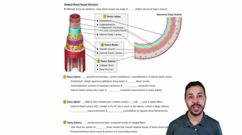
General Blood Vessel Structure
Pratica correlata
Domanda del libro di testo
1133
views
Domanda del libro di testo
Trace the normal pathway of an electrical impulse through the conducting system of the heart.
927
views
Domanda del libro di testo
What role do the chordae tendineae and papillary muscles play in the normal function of the AV valves?
1322
views
Domanda del libro di testo
During diastole, a chamber of the heart:
(a) Relaxes and fills with blood
(b) Contracts and pushes blood into an adjacent chamber
(c) Experiences a sharp increase in pressure
(d) Reaches a pressure of approximately 120 mm Hg
976
views
Domanda del libro di testo
What is the cardiac cycle? What phases and events are necessary to complete a cardiac cycle?
824
views
Domanda del libro di testo
During the cardiac cycle, the amount of blood ejected from the left ventricle when the semilunar valve opens is the:
(a) Stroke volume (SV)
(b) End-diastolic volume (EDV)
(c) End-systolic volume (ESV)
(d) Cardiac output (CO)
1192
views