Ch. 7 - DNA Structure and Replication
Sanders - Genetic Analysis: An Integrated Approach 3rd Edition
Sanders3rd EditionGenetic Analysis: An Integrated ApproachISBN: 9780135564172당신이 사용하는 게 아니라요?교과서 변경
7장, 문제 26c

Telomeres are found at the ends of eukaryotic chromosomes. What is the functional role of telomeres?

검증된 단계별 안내
1
Telomeres are repetitive nucleotide sequences located at the ends of eukaryotic chromosomes. They consist of a specific DNA sequence, such as TTAGGG in humans, repeated many times.
The primary function of telomeres is to protect the ends of chromosomes from degradation and prevent them from being recognized as broken DNA by the cell's repair machinery.
During DNA replication, the enzymes responsible for copying DNA cannot fully replicate the very ends of linear chromosomes. This is known as the 'end-replication problem.' Telomeres help mitigate this issue by acting as a buffer zone, ensuring that the loss of DNA during replication does not affect essential genes.
Telomeres also play a role in maintaining chromosomal stability by preventing the ends of chromosomes from fusing with each other, which could lead to genomic instability and cell malfunction.
Over time, telomeres shorten with each cell division, and when they become critically short, the cell may enter a state called senescence (a non-dividing state) or undergo apoptosis (programmed cell death). This process is associated with aging and the regulation of cellular lifespan.

비슷한 문제에 대한 검증된 영상 답변:

이 영상 해법은 위 문제에 도움이 된다고 튜터들이 추천한 것입니다.
영상 길이:
3m
도움이 되었나요?

주요 개념

질문에 올바르게 답하기 위해 반드시 이해해야 하는 핵심 개념들은 다음과 같습니다.

Telomeres

Telomeres are repetitive nucleotide sequences located at the ends of eukaryotic chromosomes. They protect the chromosome ends from deterioration and prevent them from fusing with neighboring chromosomes. Each time a cell divides, telomeres shorten, which is associated with aging and cellular senescence.
추천 영상:
가이드 코스
08:38
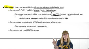
Telomeres and Telomerase

Chromosome Stability

The stability of chromosomes is crucial for maintaining genetic integrity during cell division. Telomeres play a vital role in chromosome stability by preventing the loss of essential genetic information during DNA replication. Without telomeres, chromosomes would become unstable, leading to genomic instability and potential cell death.
추천 영상:
가이드 코스
07:10
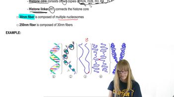
Chromosome Structure

Cellular Aging

Cellular aging, or senescence, is the process by which cells lose the ability to divide and function effectively over time. The progressive shortening of telomeres with each cell division is a key factor in this process. When telomeres become critically short, cells enter a state of senescence, contributing to aging and age-related diseases.
추천 영상:
가이드 코스
02:43
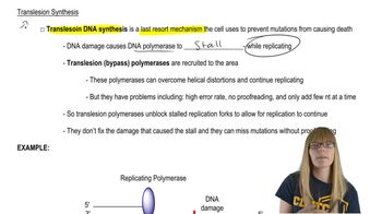
Translesion Synthesis
관련 실천
교과서 질문

Telomeres are found at the ends of eukaryotic chromosomes. What is the sequence composition of telomeres?

515
views
교과서 질문

Telomeres are found at the ends of eukaryotic chromosomes. How does telomerase assemble telomeres?

521
views
교과서 질문

A family consisting of a mother (I-1), a father (I-2), and three children (II-1, II-2, and II-3) are genotyped by PCR for a region of an autosome containing repeats of a 10-bp sequence. The mother carries 16 repeats on one chromosome and 21 on the homologous chromosome. The father carries repeat numbers of 18 and 26.

Identify all the possible genotypes of children of this couple by specifying PCR fragment lengths in each genotype.

542
views
교과서 질문

How does rolling circle replication differ from bidirectional replication?

823
views
교과서 질문

Telomeres are found at the ends of eukaryotic chromosomes. Why is telomerase usually active in germ-line cells but not in somatic cells?

860
views
교과서 질문

A family consisting of a mother (I-1), a father (I-2), and three children (II-1, II-2, and II-3) are genotyped by PCR for a region of an autosome containing repeats of a 10-bp sequence. The mother carries 16 repeats on one chromosome and 21 on the homologous chromosome. The father carries repeat numbers of 18 and 26.

Following the layout of the following figure, which aligns members of a pedigree with their DNA fragments in a gel, draw a DNA gel containing the PCR fragments generated by amplification of DNA from the parents (I-1 and I-2). Label the size of each fragment.

679
views