Ch. 19 - Pathogenic Gram-Positive Bacteria
Bauman - Microbiology with Diseases by Taxonomy 6th Edition
Bauman6th EditionMicrobiology with Diseases by TaxonomyISBN: 9780134832302당신이 사용하는 게 아니라요?교과서 변경
19장, 문제 1

Match the genera of pathogens to their appearance in stained smears: Actinomyces, Bacillus, Clostridium, Mycobacterium, Staphylococcus, Streptococcus.
(a) Methenamine silver <IMAGE>
(b) Gram <IMAGE>
(c) Gram <IMAGE>
(d) Acid fast <IMAGE>
(e) Gram <IMAGE>
(f) Gram <IMAGE>

검증된 단계별 안내
1
Step 1: Understand the staining techniques mentioned and their typical applications. Methenamine silver stain is often used to detect fungi and some bacteria with unique cell wall components. Gram stain differentiates bacteria into Gram-positive (purple) and Gram-negative (pink/red) based on cell wall structure. Acid-fast stain is used primarily for Mycobacterium species due to their waxy, mycolic acid-rich cell walls.
Step 2: Recall the general characteristics of each genus: Actinomyces are Gram-positive branching filamentous bacteria; Bacillus are large Gram-positive rods, often forming spores; Clostridium are Gram-positive rods, anaerobic and spore-forming; Mycobacterium are acid-fast rods; Staphylococcus are Gram-positive cocci in clusters; Streptococcus are Gram-positive cocci in chains.
Step 3: Match the genera to the staining method and expected morphology: (a) Methenamine silver stain likely highlights Actinomyces due to their filamentous nature; (b), (c), (e), and (f) are Gram stains showing different morphologies—identify which images show rods or cocci and their arrangements to assign Bacillus, Clostridium, Staphylococcus, and Streptococcus accordingly; (d) Acid-fast stain corresponds to Mycobacterium.
Step 4: Use the morphology clues from the images (if available) to distinguish between rods and cocci, and their arrangements (chains vs clusters), which helps differentiate Bacillus, Clostridium, Staphylococcus, and Streptococcus in Gram stains.
Step 5: Finalize the matching by pairing each genus with the staining method and morphology that best fits their known characteristics, ensuring Mycobacterium is matched with acid-fast stain and Actinomyces with methenamine silver stain.

비슷한 문제에 대한 검증된 영상 답변:

이 영상 해법은 위 문제에 도움이 된다고 튜터들이 추천한 것입니다.
영상 길이:
2m

주요 개념

질문에 올바르게 답하기 위해 반드시 이해해야 하는 핵심 개념들은 다음과 같습니다.

Bacterial Staining Techniques

Staining methods like Gram stain, acid-fast stain, and methenamine silver stain are used to visualize bacteria under a microscope. Each technique highlights different bacterial features based on cell wall properties, aiding in identification. For example, Gram stain differentiates bacteria into Gram-positive or Gram-negative, while acid-fast stain detects mycobacteria with waxy cell walls.
추천 영상:
02:10
Introduction to Staining

Morphology and Characteristics of Bacterial Genera

Different bacterial genera have distinct shapes and staining properties. Bacillus and Clostridium are Gram-positive rods, Actinomyces are filamentous Gram-positive bacteria, Staphylococcus and Streptococcus are Gram-positive cocci with different arrangements, and Mycobacterium is acid-fast due to mycolic acid in its cell wall. Recognizing these traits helps match bacteria to their stained appearances.
추천 영상:
02:44
Types of Bacterial Cell Morphology

Correlation Between Staining Results and Bacterial Identification

Interpreting stained smears requires linking staining patterns to bacterial identity. For instance, acid-fast positive staining indicates Mycobacterium, while methenamine silver stain is used for certain fungi and bacteria with specific cell wall components. Understanding these correlations enables accurate matching of genera to their microscopic images.
추천 영상:
05:30
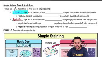
Simple Staining Basic & Acidic Dyes