Immersion oil ________(increases/decreases) the numerical aperture, which ________(increases/decreases) resolution because _______(more/fewer) light rays are involved.
Ch. 4 - Microscopy, Staining, and Classification
4장, 문제 2
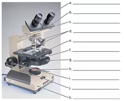
Label the microscope.

검증된 단계별 안내1
Identify the main parts of the microscope that need labeling, such as the eyepiece (ocular lens), objective lenses, stage, light source, diaphragm, coarse focus knob, fine focus knob, and arm.
Start with the eyepiece, which is the lens at the top that you look through to see the specimen. Label it as 'Eyepiece (Ocular Lens)'.
Locate the objective lenses, which are the multiple lenses attached to the rotating nosepiece just above the stage. Label each objective lens according to its magnification (e.g., 4x, 10x, 40x).
Find the stage, the flat platform where the slide is placed. Label it as 'Stage'. Also, identify the stage clips that hold the slide in place.
Identify the light source located beneath the stage, which illuminates the specimen. Label it as 'Light Source'. Then, locate the diaphragm or iris, which controls the amount of light passing through the specimen, and label it accordingly.

비슷한 문제에 대한 검증된 영상 답변:
이 영상 해법은 위 문제에 도움이 된다고 튜터들이 추천한 것입니다.
영상 길이:
2m도움이 되었나요?
주요 개념
질문에 올바르게 답하기 위해 반드시 이해해야 하는 핵심 개념들은 다음과 같습니다.
Parts of a Light Microscope
A light microscope consists of key components such as the eyepiece (ocular lens), objective lenses, stage, light source, diaphragm, and focusing knobs. Each part plays a specific role in magnifying and illuminating the specimen for observation.
추천 영상:
가이드 코스
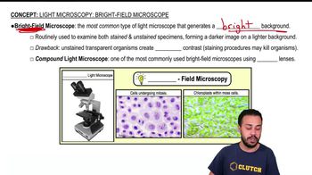
Light Microscopy: Bright-Field Microscopes
Function of Microscope Components
Understanding the function of each part is essential; for example, objective lenses provide different magnifications, the stage holds the slide, and the diaphragm controls light intensity. Proper use of focusing knobs adjusts clarity and sharpness.
추천 영상:
가이드 코스
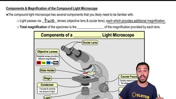
Components & Magnification of the Compound Light Microscope
Microscope Handling and Usage
Correct handling involves proper slide placement, adjusting light and focus gradually, and knowing how to switch between objective lenses. This ensures clear visualization of microorganisms or cells without damaging the equipment.
추천 영상:
가이드 코스
Light Microscopy: Bright-Field Microscopes
관련 실천
교과서 질문
1539
views
교과서 질문
A nanometer is _______ than a micrometer.
a. 10 times larger
b. 10 times smaller
c. 1000 times larger
d. 1000 times smaller
1609
views
교과서 질문
Label each photograph with the type of microscope used to acquire the image.
a. _______<IMAGE>
b. _______<IMAGE>
c. _______<IMAGE>
d. _______<IMAGE>
e. _______<IMAGE>
f. _______<IMAGE>
1512
views
교과서 질문
The type of fixation developed by Koch for bacteria is _______.
1859
views
교과서 질문
Resolution is best described as the ____________.
a. Ability to view something that is small
b. Ability to magnify a specimen
c. Ability to distinguish between two adjacent objects
d. Difference between two waves of electromagnetic radiation
1536
views
교과서 질문
Critique the following definition of magnification given by a student on a microbiology test: “Magnification makes things bigger.”
1678
views