Ch. 4 - Microscopy, Staining, and Classification
Bauman - Microbiology with Diseases by Taxonomy 6th Edition
Bauman6th EditionMicrobiology with Diseases by TaxonomyISBN: 9780134832302당신이 사용하는 게 아니라요?교과서 변경
4장, 문제 1

Label each photograph with the type of microscope used to acquire the image.


a. _______<IMAGE>
b. _______<IMAGE>
c. _______<IMAGE>
d. _______<IMAGE>
e. _______<IMAGE>
f. _______<IMAGE>

검증된 단계별 안내
1
Step 1: Review the characteristics of each type of microscope commonly used in microbiology, such as light microscopes (bright-field, phase-contrast, fluorescence), electron microscopes (scanning electron microscope - SEM, transmission electron microscope - TEM), and confocal microscopes.
Step 2: Examine each photograph carefully, noting key features such as color, resolution, depth of field, and the type of detail visible (surface texture, internal structures, fluorescence patterns).
Step 3: Match the observed features in each image to the known capabilities of each microscope type. For example, bright-field microscopy shows stained cells with color, SEM provides detailed 3D surface images, TEM shows high-resolution internal structures in black and white, and fluorescence microscopy shows bright colors against a dark background.
Step 4: Label each photograph with the microscope type that best fits the observed image characteristics based on your analysis from Step 3.
Step 5: Double-check your labels by comparing them to reference images or descriptions from your textbook or lecture notes to ensure accuracy.

비슷한 문제에 대한 검증된 영상 답변:

이 영상 해법은 위 문제에 도움이 된다고 튜터들이 추천한 것입니다.
영상 길이:
2m
도움이 되었나요?

주요 개념

질문에 올바르게 답하기 위해 반드시 이해해야 하는 핵심 개념들은 다음과 같습니다.

Types of Microscopes

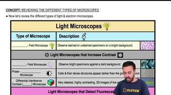
Microscopes vary based on their imaging techniques and magnification capabilities, including light microscopes (bright-field, phase-contrast, fluorescence) and electron microscopes (scanning and transmission). Each type is suited for different sample types and resolutions, influencing the image's appearance.
추천 영상:
가이드 코스
05:32
Reviewing the Different Types of Microscopes

Image Characteristics and Resolution

Different microscopes produce images with distinct features such as color, contrast, depth, and resolution. For example, electron microscopes provide high-resolution, black-and-white images showing fine structural details, while light microscopes often show colored or stained samples with lower resolution.
추천 영상:
가이드 코스
07:29
Magnification, Resolution, & Contrast

Sample Preparation Techniques

The method used to prepare samples affects the microscope type and image quality. Techniques like staining, freezing, or coating with metals are specific to certain microscopes and help enhance contrast or preserve structures, aiding in correct identification of the microscope used.
추천 영상:
가이드 코스
02:05
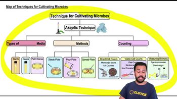
Map of Techniques for Cultivating Microbes