Microbiology
My Course
Learn
Exam Prep
AI Tutor
Study Guides
Textbook Solutions
Flashcards
Explore
Try the app
My Course
Learn
Exam Prep
AI Tutor
Study Guides
Textbook Solutions
Flashcards
Explore
Try the app
Back
Introduction to Light Microscopy definitions
You can tap to flip the card.
Light Microscopy
You can tap to flip the card.
👆
Light Microscopy
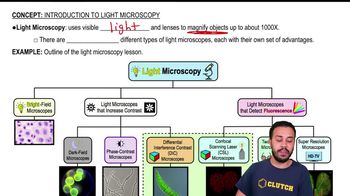
Technique using visible light and lenses to magnify small objects, enabling visualization up to 1,000 times their actual size.
Track progress
Control buttons has been changed to "navigation" mode.
1/14
Related flashcards
Related practice
Recommended videos
Introduction to Light Microscopy quiz #1
Introduction to Light Microscopy
10 Terms
Introduction to Light Microscopy
9. Microscopes
1 problem
Topic
Light Microscopy: Bright-Field Microscopes
9. Microscopes
4 problems
Topic

Brendan
9. Microscopes - Part 1 of 2
6 topics
12 problems
Chapter
Nicole
9. Microscopes - Part 2 of 2
8 topics
11 problems
Chapter
Nicole
Guided course
02:53
Introduction to Light Microscopy
2581
views
36
rank
Terms in this set (14)
Hide definitions
Light Microscopy
Technique using visible light and lenses to magnify small objects, enabling visualization up to 1,000 times their actual size.
Magnification
Increase in apparent size of an object, allowing detailed observation of structures too small for the naked eye.
Bright Field Microscope
Instrument that illuminates specimens with visible light, producing a bright background for observing stained or naturally pigmented samples.
Dark Field Microscope
Device enhancing contrast by blocking direct light, making specimens appear bright against a dark background, ideal for transparent samples.
Phase Contrast Microscope
Tool that amplifies differences in refractive index, allowing visualization of transparent specimens without staining.
Differential Interference Contrast Microscope
Instrument using polarized light to create high-contrast, three-dimensional-like images of unstained cells.
DIC Microscope
Specialized device for imaging cells in three-dimensional space, providing enhanced contrast and depth.
Fluorescence Detection
Method for visualizing specimens by detecting emitted light from fluorescent molecules, revealing specific structures.
Confocal Scanning Laser Microscope
Advanced system using lasers to scan specimens, producing sharp, three-dimensional images by eliminating out-of-focus light.
CSL Microscope
Instrument crucial for three-dimensional imaging of cells, utilizing laser scanning and fluorescence.
2 Photon Microscope
Device enabling deep tissue imaging by using two photons of lower energy, minimizing damage to living samples.
Super Resolution Microscope
Technology surpassing traditional optical limits, allowing visualization of structures at the nanometer scale.
Contrast
Difference in light intensity between specimen and background, essential for distinguishing details in microscopy.
Three-Dimensional Imaging
Visualization technique providing spatial information about cellular structures, enhancing depth perception.