Skip to main content
Microbiology
My Course
Learn
Exam Prep
AI Tutor
Study Guides
Textbook Solutions
Flashcards
Explore
Try the app
My Course
Learn
Exam Prep
AI Tutor
Study Guides
Textbook Solutions
Flashcards
Explore
Try the app
Back
Light Microscopes that Increase Contrast definitions
You can tap to flip the card.
Bright Field Microscope
You can tap to flip the card.
👆
Bright Field Microscope
Instrument producing images with a dark specimen on a bright background, often requiring staining for better visibility.
Track progress
Control buttons has been changed to "navigation" mode.
1/14
Related flashcards
Related practice
Recommended videos
Light Microscopes that Increase Contrast quiz
Light Microscopes that Increase Contrast
15 Terms
Light Microscopes that Increase Contrast
9. Microscopes
2 problems
Topic
Nicole
Light Microscopes that Detect Fluorescence
9. Microscopes
5 problems
Topic
Monica
9. Microscopes - Part 1 of 2
6 topics
12 problems
Chapter
Nicole
9. Microscopes - Part 2 of 2
8 topics
10 problems
Chapter
Nicole
Guided course
02:39
Light Microscopes that Increase Contrast
2187
views
40
rank
Guided course
01:52
Dark-Field Microscopy
1911
views
25
rank
Guided course
03:52
Phase-Contrast Microscopy
2088
views
33
rank
Terms in this set (14)
Hide definitions
Bright Field Microscope
Instrument producing images with a dark specimen on a bright background, often requiring staining for better visibility.
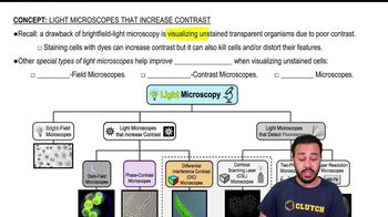
Contrast
Degree of visual difference between a specimen and its background, crucial for distinguishing cell features.
Staining
Process using dyes to color cells, enhancing visibility but potentially killing or distorting specimens.
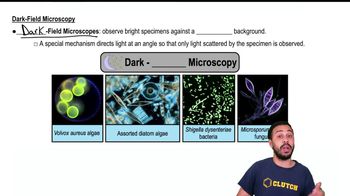
Dark Field Microscope
Instrument creating images with a bright specimen on a dark background by detecting only scattered light.
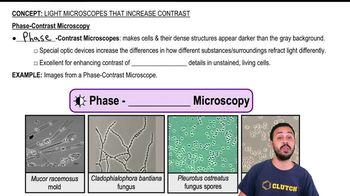
Phase Contrast Microscope
Device using special optics to amplify light refraction differences, revealing internal details in unstained living cells.
Differential Interference Contrast Microscope
Instrument generating highly detailed, three-dimensional images with added color contrast using advanced optics.
Optic Devices
Complex components within microscopes that manipulate light to enhance image contrast and reveal fine details.
Background
Visual field surrounding the specimen, which can be manipulated to improve specimen visibility in microscopy.
Three-dimensional Image
Visual output providing depth and detail, making specimens appear to extend off the viewing surface.
Unstained Cell
Living specimen observed without dyes, maintaining natural features but often requiring special microscopy for visibility.
Internal Structure
Cellular components and organelles made visible by advanced contrast techniques without the need for staining.
Scattered Light
Illumination redirected by a specimen, used in certain microscopes to enhance the specimen's visibility.
Grayish Background
Neutral field often produced in phase contrast microscopy, making cells and structures appear darker for better contrast.
Live Specimen
Organism or cell observed in its natural, undisturbed state, often requiring non-destructive imaging methods.
 BackBack
 02:39
02:39