Skip to main content
Microbiology
My Course
Learn
Exam Prep
AI Tutor
Study Guides
Textbook Solutions
Flashcards
Explore
Try the app
My Course
Learn
Exam Prep
AI Tutor
Study Guides
Textbook Solutions
Flashcards
Explore
Try the app
Back
Other Types of Staining definitions
You can tap to flip the card.
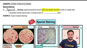
Special Staining
You can tap to flip the card.
👆
Special Staining
Technique using unique procedures to highlight specific internal or external cell structures for easier visualization.
Track progress
Control buttons has been changed to "navigation" mode.
1/13
Related flashcards
Related practice
Recommended videos
Other Types of Staining quiz
Other Types of Staining
15 Terms
Other Types of Staining
9. Microscopes
3 problems
Topic
Nicole
9. Microscopes - Part 1 of 2
6 topics
12 problems
Chapter
Nicole
9. Microscopes - Part 2 of 2
8 topics
10 problems
Chapter
Nicole
Guided course
02:59
Fluorescent Dyes
1745
views
18
rank
Guided course
02:59
Special Staining
2156
views
21
rank
Terms in this set (13)
Hide definitions
Special Staining
Technique using unique procedures to highlight specific internal or external cell structures for easier visualization.
Capsule Stain
Method that reveals the protective barrier around bacteria as a white border, making it visible under the microscope.
Endospore Stain
Procedure that colors dormant bacterial structures bluish-green, aiding in their identification within cells.
Flagella Stain
Technique that makes thin, whip-like appendages of bacteria visible, which are otherwise undetectable.
Capsule
Gel-like outer layer surrounding some bacteria, seen as a white border when stained, providing protection.
Endospore
Highly resistant, dormant structure formed by certain bacteria, visible as blue-green dots after special staining.
Flagella
Long, thread-like structures extending from bacteria, responsible for movement, visualized only with specific stains.
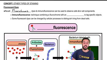
Fluorescent Dye
Chemical that emits visible light when excited, used to tag and visualize cellular components under special microscopes.
Immunofluorescence
Technique combining fluorescent dyes with antibodies to detect specific molecules on or within cells.
Antibody
Y-shaped protein that binds specifically to target molecules, enabling their detection when linked to fluorescent dyes.
Antigen
Molecule recognized and bound by antibodies, allowing for targeted visualization in immunofluorescence.
Fluorochrome
Type of fluorescent dye that emits light upon excitation, often attached to antibodies for cell labeling.
Cellular Process
Biochemical activity within cells that can alter certain fluorescent dyes, distinguishing living from dead cells.
 BackBack
 02:59
02:59