 Back
Bacteriophages and Virus Basics - Microbiology
 04:47

Terms in this set (15)
Living: Can reproduce only inside host cells. Non-living: No metabolism, no cellular structure, cannot reproduce independently.
Viruses are much smaller than bacteria, typically 20-300 nm, while bacteria are about 1,000 nm (1 µm) or larger.
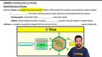
Viruses consist of a nucleic acid core surrounded by a protein coat called a capsid. The capsid is made of protein subunits called capsomers. The nucleic acid plus capsid is the nucleocapsid.
Viruses contain either DNA or RNA, which can be single-stranded or double-stranded, but never both.
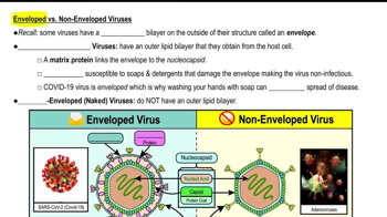
Naked viruses have only a capsid and nucleic acid. Enveloped viruses have an outer lipid membrane derived from the host cell.
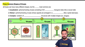
Bacteriophages are viruses that infect bacteria. They have complex structures and replicate via the lytic or lysogenic cycle.
Bacteriophages attach to bacteria, inject nucleic acid, replicate inside, and often lyse the host cell to release new phages (lytic cycle).
Phage typing is a method to identify bacterial strains based on their susceptibility to specific bacteriophages.
Phage specificity is shown by clear zones of lysis on bacterial lawns where only susceptible bacteria are killed by a particular phage.
Sensitive bacteria show clear plaques (zones of lysis). Insensitive bacteria show no plaques and grow normally.
Spread bacteria on agar, apply different phages, incubate, and observe plaques to determine bacterial susceptibility.
A plaque is a clear zone on a bacterial lawn where phages have lysed bacterial cells.
Look for small, clear circular areas on the otherwise opaque bacterial lawn indicating phage-induced lysis.
Count the number of plaques on a plate to estimate the number of infectious phage particles in the sample.
PFUs are calculated by multiplying the number of plaques by the dilution factor: \(\text{PFU/mL} = \frac{\text{Number of plaques}}{\text{Dilution} \times \text{Volume plated}}\)