 Back
Microbiology: Immune Deficiencies, Autoimmunity, and Hypersensitivities
 02:41

Terms in this set (46)
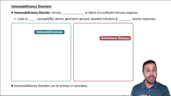
Immunodeficiency is a condition where the immune system's ability to fight infectious disease is compromised or entirely absent.
Primary immunodeficiencies are congenital disorders present from birth that result in a defective immune system.
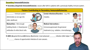
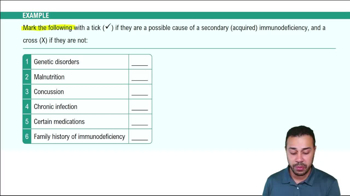
Secondary immunodeficiencies develop after birth due to external factors like infections, malnutrition, or medical treatments.
Viral infections are the leading cause of secondary immunodeficiencies besides aging.
HIV, measles virus, cytomegalovirus, and Epstein-Barr virus can cause secondary immunodeficiencies.
Kaposi sarcoma is a cancer of blood vessel walls; late-stage HIV infection increases susceptibility.
Prophylactic immunotherapy prevents disease; therapeutic immunotherapy treats existing disease.
An autoimmune disorder occurs when the immune system mistakenly attacks the body’s own tissues.
Failure of self-tolerance causes the immune system to attack self-antigens, leading to autoimmune disorders.
Type I diabetes: Coxsackievirus; Guillain–Barré syndrome: Campylobacter jejuni; Rheumatic heart disease: Streptococcus pyogenes; Multiple sclerosis: Epstein-Barr virus.
False. Autoimmune diseases often require multiple tests and complex diagnosis.
Hypersensitivities are caused by an exaggerated or inappropriate immune response to an antigen.
The hygiene hypothesis suggests reduced early-life exposure to microbes increases susceptibility to allergies and autoimmune diseases.
The Gell and Coombs classification organizes hypersensitivities into Types I, II, III, and IV.
Type IV hypersensitivity is cell-mediated and not antibody-mediated.
An allergen is a harmless antigen causing an immune response; allergy is the hypersensitive reaction to it.
Sensitizing exposure is the first contact with an allergen that induces IgE production without symptoms.
Mast cells and basophils degranulate, releasing histamine, leukotrienes, and prostaglandins.
Localized anaphylaxis affects a specific area; systemic anaphylaxis affects the whole body and can cause shock.
Anaphylactic shock is a severe systemic allergic reaction treated with epinephrine.
Increased IgE indicates sensitization to a specific allergen.
A wheal and flare is a raised, red, itchy area indicating a positive allergic reaction to the tested allergen.
To reduce allergic sensitivity by gradually increasing allergen exposure to shift immune response away from IgE.
IgG and IgM antibodies bind to antigens on cell surfaces or extracellular matrix.
Antibodies block receptor function causing altered cell activity; outcomes include cell dysfunction or death.
Type II hypersensitivity includes transfusion reactions due to blood group incompatibility.
Both A and B antigens are present; Rh antigen is absent.
Recipient antibodies attack donor RBC antigens, causing hemolysis and possible severe complications.
Anti-B antibodies in recipient attack B antigens on transfused RBCs, causing hemolysis.
AB+ is universal recipient because it has all A, B, and Rh antigens and no anti-A or anti-B antibodies.
O- is universal donor because it lacks A, B, and Rh antigens, minimizing immune reaction.
Mother is sensitized but no hemolytic disease occurs in first pregnancy.
Maternal anti-Rh antibodies cross placenta and cause hemolytic disease of the newborn.
Rho(D) immune globulin (RhoGAM) prevents sensitization to Rh antigen.
IgG and IgM antibodies bind to soluble antigens forming immune complexes.
Immune complexes deposit in tissues causing inflammation and tissue damage.
Lupus, rheumatoid arthritis, serum sickness, glomerulonephritis, and Arthus reaction.
After injection of foreign serum, immune complexes form and deposit causing systemic inflammation.
T cells, especially CD4+ and CD8+ T cells, mediate Type IV hypersensitivity.
Because symptoms appear 24-72 hours after antigen exposure due to T cell activation.
Type 1 diabetes, multiple sclerosis, rheumatoid arthritis, Hashimoto thyroiditis, and Crohn’s disease.
Haptens, small molecules that bind to proteins and trigger T cell response.
Contact dermatitis, tuberculin skin test, graft rejection, and chronic transplant rejection.
Autograft: self to self; Isograft: identical twin; Allograft: same species; Xenograft: different species. Autograft least likely rejected.
Sites where immune response is limited: brain, eyes, testes, and placenta.
Transplant rejection: host attacks graft; Graft-vs-host: graft immune cells attack host tissues.