 Back
BackA Brief History of Microbiology and Microscopy: Foundations, Key Discoveries, and Laboratory Techniques
Study Guide - Smart Notes
 Tailored notes based on your materials, expanded with key definitions, examples, and context.
Introduction to Microbiology
Definition and Scope
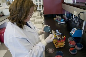
Microbiology is the scientific study of organisms too small to be seen with the naked eye, including bacteria, archaea, fungi, protozoa, helminths, viruses, and prions. This field explores their structure, function, classification, and roles in health, disease, and the environment.
Microbes are ubiquitous and play essential roles in ecosystems, industry, and medicine.
Microbiology integrates concepts from biology, chemistry, and physics to understand microscopic life.
Major Groups of Microorganisms
Bacteria and Archaea
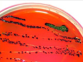
Bacteria and archaea are unicellular prokaryotes lacking a nucleus. They are found in diverse environments, including extreme habitats, and reproduce asexually.
Bacteria: Common in soil, water, and living hosts; some are pathogenic.
Archaea: Often inhabit extreme environments (e.g., hot springs, salt lakes).

Fungi
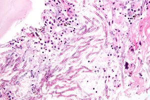
Fungi are eukaryotic organisms that obtain nutrients from other organisms and possess cell walls. They include multicellular molds and unicellular yeasts.
Molds: Multicellular, form hyphae, reproduce by spores.
Yeasts: Unicellular, reproduce by budding or spores.
Protozoa (Protists)
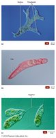
Protozoa are single-celled eukaryotes with animal-like characteristics. They are mostly aquatic and exhibit various forms of locomotion.
Reproduce asexually (most) or sexually.
Some are free-living; others are parasitic.
Helminths
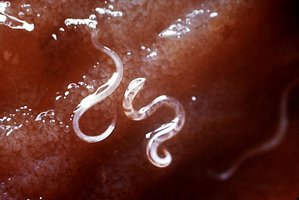
Helminths are parasitic worms, including tapeworms and roundworms. While adult forms are macroscopic, their eggs and larvae are microscopic and studied in microbiology.
Multicellular eukaryotes.
Cause significant human and animal diseases.
Acellular Infectious Agents: Viruses and Prions
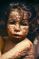
Viruses are acellular entities that require host cells for replication. Prions are infectious proteins lacking genetic material.
Viruses: Cause a wide range of diseases; much smaller than bacteria.
Prions: Cause neurodegenerative diseases (e.g., mad cow disease, CJD).
Historical Foundations of Microbiology
Spontaneous Generation vs. Biogenesis
Early scientists debated whether life could arise spontaneously (abiogenesis) or only from pre-existing life (biogenesis).
Spontaneous Generation: Proposed by Aristotle; life arises from non-living matter.
Biogenesis: Life arises only from existing life; supported by later experiments.
Redi’s Experiments
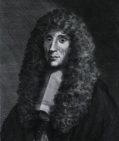
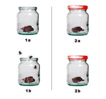
Francesco Redi demonstrated that maggots on meat arose from flies, not spontaneous generation, by isolating meat from flies in sealed containers.
Helped disprove abiogenesis for larger organisms.
Antonie van Leeuwenhoek and Robert Hooke
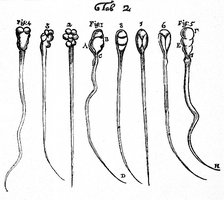
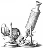
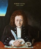
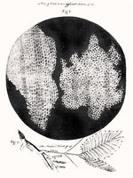
Antonie van Leeuwenhoek developed powerful microscopes and was the first to observe and describe bacteria and protozoa. Robert Hooke coined the term "cell" and contributed to cell theory.
Leeuwenhoek: "Father of Microbiology"; discovered "animalcules" (microbes).
Hooke: First described cells in cork; published first microorganism description.

Louis Pasteur and the Germ Theory of Disease
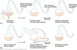
Louis Pasteur disproved spontaneous generation for microbes using swan-neck flask experiments. He established that microbes cause fermentation and spoilage, leading to the germ theory of disease and the development of pasteurization and vaccines.
Demonstrated that microbes do not arise from non-living matter.
Developed vaccines for rabies and anthrax.
Robert Koch and Koch’s Postulates
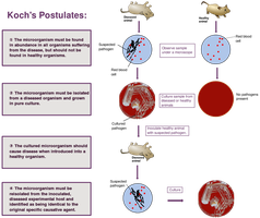
Robert Koch established methods to link specific microbes to specific diseases, formulating Koch’s postulates. He isolated important pathogens and advanced laboratory techniques.
Isolated Bacillus anthracis, Mycobacterium tuberculosis, and Vibrio cholerae.
Developed pure culture techniques and staining methods.
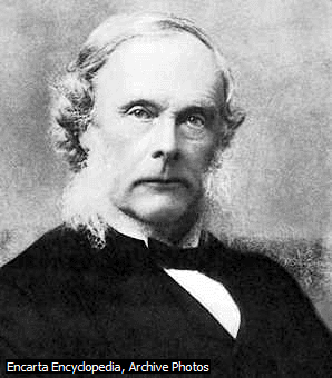
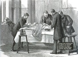
Joseph Lister and Antiseptic Technique
Joseph Lister introduced antiseptic techniques in surgery, drastically reducing infections by sterilizing instruments and using disinfectants.
Promoted handwashing, sterilization, and use of carbolic acid.
Ignaz Semmelweis and Handwashing
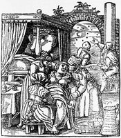
Ignaz Semmelweis demonstrated that handwashing in obstetrical clinics reduced puerperal fever, highlighting the importance of hygiene in preventing disease transmission.
Microscopy and Microbiological Tools
Microscopy: Principles and Types
Microscopy is essential for visualizing microbes. Key concepts include wavelength, magnification, contrast, and resolution. The main types of microscopes are light (simple, compound) and electron (SEM, TEM).
Magnification: Enlargement of an image.
Resolution: Ability to distinguish two points as separate.
Contrast: Difference in light intensity between specimen and background.
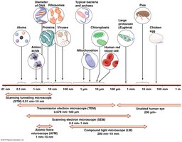
Types of Microscopes
Bright-field: Most common; uses visible light.
Electron Microscopes: Use electron beams for much higher magnification and resolution.
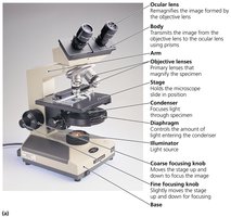
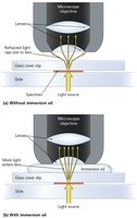
Immersion Oil and Resolution
Immersion oil increases resolution by reducing light refraction between the specimen and the objective lens.
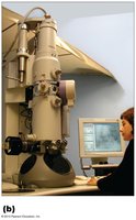
Electron Microscopy
Electron microscopes allow visualization of cell ultrastructure at magnifications up to 100,000x. Two main types are:
Transmission Electron Microscope (TEM): Views internal structures.
Scanning Electron Microscope (SEM): Views surface details.
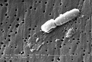
Staining and Microbial Morphology
Staining Techniques
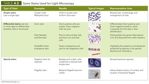
Staining enhances contrast and reveals cellular structures. Stains are classified as simple, differential, or special.
Simple stains: Single dye; reveals cell shape and arrangement.
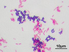
Differential stains: Multiple dyes; distinguishes cell types (e.g., Gram stain, acid-fast stain).
Special stains: Highlight specific structures (e.g., capsule, flagella).
Type of Stain | Examples | Results | Typical Images | Representative Uses |
|---|---|---|---|---|
Simple stain | Crystal violet, Methylene blue | Uniform purple/blue cells | Cell morphology and arrangement | Reveals cell shape, arrangement |
Differential stain | Gram stain, Acid-fast stain | Gram-positive (purple), Gram-negative (pink); Acid-fast (pink), Non-acid-fast (blue) | Bacterial classification | Distinguishes bacteria, detects pathogens |
Special stain | Negative stain, Flagellar stain | Capsules appear as clear halos; flagella visible | Specific structures | Identifies capsules, flagella |
Microbial Morphology
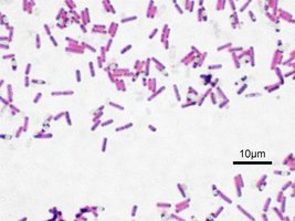
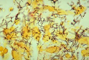
Bacteria exhibit characteristic shapes and arrangements, which aid in identification.
Coccus: Spherical
Bacillus: Rod-shaped
Spirillum: Spiral-shaped
Pleomorphism: Variation in shape/size within a species
Summary and Key Concepts
Microbiology studies diverse microscopic life forms and their impact on health and the environment.
Key historical experiments disproved spontaneous generation and established the germ theory of disease.
Microscopy and staining are essential tools for observing and classifying microbes.
Understanding microbial morphology and laboratory techniques is foundational for further study in microbiology.
