 Back
BackAcellular Infectious Agents: Viruses, Prions, and Viroids – Structure, Classification, and Life Cycles
Study Guide - Smart Notes
 Tailored notes based on your materials, expanded with key definitions, examples, and context.
Acellular Infectious Agents
Overview of Acellular Infectious Agents
Acellular infectious agents are pathogens that lack cellular structure and cannot replicate independently. They include viruses, prions, and viroids. Each type has distinct structural and functional characteristics, but all require a living host cell for replication.
Viruses: Infectious particles composed of a nucleic acid genome (DNA or RNA) encased in a protein coat (capsid); some possess a lipid envelope.
Prions: Infectious proteins that lack nucleic acids and cause neurodegenerative diseases.
Viroids: Infectious, small, circular RNA molecules without a protein coat, primarily infecting plants.
All acellular agents depend on host cells for replication and can infect a wide range of organisms, including bacteria, fungi, plants, and animals.
Viral Structure
Virion Components and Organization
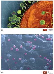
The extracellular form of a virus is called a virion. Virions are composed of a nucleic acid genome and a protein coat called a capsid. Some viruses also possess a lipid envelope derived from the host cell membrane.
Nucleocapsid: The combination of the viral genome and capsid.
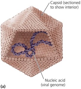
Capsid: Made of protein subunits called capsomeres; protects the genome and determines the virus's shape.
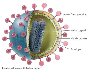
Envelope: A lipid bilayer surrounding some nucleocapsids, containing both host and viral proteins (e.g., glycoproteins) that facilitate host recognition and attachment.
Capsid Shapes
Capsids can take on several distinct shapes, which are important for virus classification and function.
Helical: Rod-shaped structure formed by capsomeres arranged in a spiral around the genome.
Polyhedral (Icosahedral): Nearly spherical, with 20 triangular faces; common among animal viruses.
Complex: Structures that do not fit into the helical or polyhedral categories, such as bacteriophages with heads and tails.
Viral Envelopes
Enveloped viruses have a lipid bilayer surrounding their nucleocapsid, which provides protection and aids in host cell recognition. The envelope contains viral glycoproteins and host-derived proteins. Non-enveloped (naked) viruses lack this lipid layer and consist only of the nucleocapsid.
Viral Genomes and Classification
Types of Viral Genomes
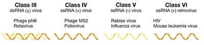
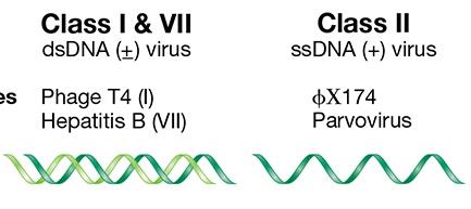
Viral genomes are highly variable and can be composed of DNA or RNA, which may be single-stranded (ss) or double-stranded (ds). The nature of the genome determines the replication strategy and classification of the virus.
DNA Viruses: Can be dsDNA or ssDNA.
RNA Viruses: Can be dsRNA or ssRNA. ssRNA viruses are further classified as plus-sense (+ssRNA) (same sequence as mRNA) or minus-sense (-ssRNA) (complementary to mRNA).
The Baltimore classification system groups viruses based on genome type and replication mechanism.
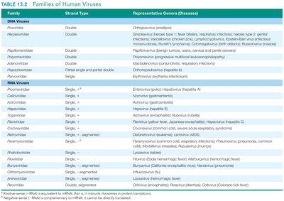
Examples of Human Virus Genomes
Human viruses display a wide range of genome types, which are summarized in the following table:
Family | Strand Type | Representative Genera (Diseases) |
|---|---|---|
Parvoviridae | Single, - | Erythrovirus (fifth disease) |
Herpesviridae | Double | Herpes simplex virus (oral/genital herpes), Varicella-zoster virus (chickenpox) |
Picornaviridae | Single, + | Enterovirus (poliomyelitis), Rhinovirus (common cold) |
Orthomyxoviridae | Single, -, segmented | Influenzavirus (influenza) |
Retroviridae | Single, +, two copies, uses DNA intermediate | Lentivirus (AIDS) |
Viral Hosts and Host Specificity
Host Range and Specificity
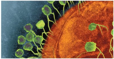
Viruses can infect all forms of life, including bacteria (bacteriophages), archaea, fungi, plants, and animals. Host specificity is determined by the ability of the virion to attach to specific receptors on the host cell surface. Some viruses are generalists, infecting multiple hosts, while others are highly specific.
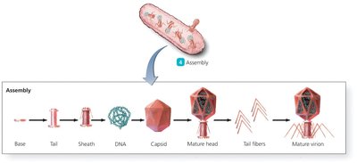
Bacteriophage Structure
Unique Features of Bacteriophages
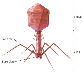
Bacteriophages (phages) are viruses that infect bacteria. They often have complex structures with a distinct head (nucleocapsid) and tail apparatus used for attachment and genome injection.
Head: Contains the viral genome and may carry enzymes.
Tail: Includes tail fibers for host recognition, a tail tube for genome delivery, and a base plate for attachment.
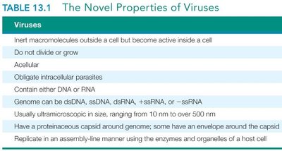
Unique Features of Viruses vs. Cells
Comparison Table: Cells vs. Viruses
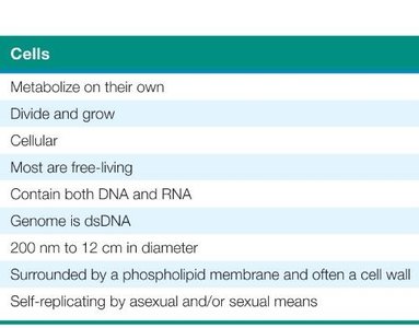
Viruses differ fundamentally from cells in structure, replication, and metabolism. The following table summarizes these differences:
Feature | Cells | Viruses |
|---|---|---|
Metabolism | Yes | No |
Growth & Division | Yes | No |
Cellular Structure | Yes | No (acellular) |
Genome | DNA & RNA | DNA or RNA |
Replication | Self-replicating | Obligate intracellular parasite |
Size | 200 nm – 12 cm | 10 nm – 500 nm |
Membrane | Phospholipid membrane, often cell wall | Protein capsid, sometimes envelope |
Viral Life Cycle
General Steps of Viral Replication
The viral life cycle consists of five main steps, each critical for the production of new virions:
Attachment (Adsorption): Virion binds to specific receptors on the host cell surface.
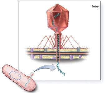
Entry: The virion or its genome enters the host cell. For bacteriophages, only the genome enters, while the capsid remains outside.
Synthesis: Host cell machinery is redirected to synthesize viral nucleic acids and proteins.
Assembly: New virions are assembled from synthesized components.
Release: Mature virions exit the host cell, either by lysis (cell destruction) or budding (enveloped viruses).
Attachment and Entry
Attachment is mediated by interactions between viral proteins (capsid or envelope glycoproteins) and host cell receptors. Entry mechanisms vary:
Enveloped viruses may fuse with the host membrane or enter via endocytosis.
Bacteriophages inject their genome through the bacterial cell wall using specialized structures and enzymes (e.g., lysozyme).
Synthesis, Assembly, and Release
Once inside, the viral genome directs the host cell to produce viral components. Assembly is a highly ordered process, and release occurs by:
Lysis: Host cell bursts, releasing all virions simultaneously (common in bacteriophages).
Budding: Virions acquire an envelope as they exit the host cell (common in animal viruses).
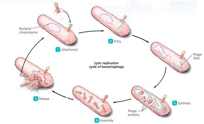
Lytic and Lysogenic Cycles in Bacteriophages
Lytic Cycle
The lytic cycle is characterized by the active replication of the phage, culminating in host cell lysis and release of new virions. The steps include attachment, entry, synthesis, assembly, and release.
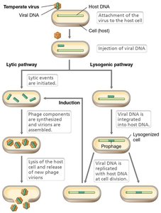
Lysogenic Cycle (Temperate Phages)
Temperate phages can integrate their genome into the host chromosome, becoming a prophage. The viral genome is replicated along with the host cell's DNA and can remain dormant until induced to enter the lytic cycle.
Induction: Environmental signals can trigger the prophage to excise from the host genome and initiate the lytic cycle.

Key Concepts and Review Questions
Why can’t all viruses infect all kinds of cells? Viruses require specific receptors on host cells for attachment; only cells with compatible receptors can be infected.
What happens if a phage is missing its tail fibers? Attachment to the host cell is disrupted, preventing infection.
Why doesn’t the entire virion enter a bacterial cell? The bacterial cell wall is rigid and only allows the viral genome to be injected, not the whole capsid.
Summary Table: Acellular Infectious Agents
Agent | Structure | Genome | Host Range |
|---|---|---|---|
Virus | Protein capsid, sometimes envelope | DNA or RNA | Bacteria, Archaea, Eukaryotes |
Prion | Protein only | None | Animals (mainly) |
Viroid | RNA only | ssRNA | Plants |
