 Back
BackAcellular Infectious Agents: Viruses, Viroids, and Prions – Structure, Life Cycles, and Pathogenesis
Study Guide - Smart Notes
 Tailored notes based on your materials, expanded with key definitions, examples, and context.
Viruses: Structure and Life Cycle
General Virus Life Cycle
Viruses are acellular infectious agents that require host cells for replication. The viral life cycle consists of five main steps:
Attachment (Adsorption): The virion binds to specific receptors on the host cell surface.
Entry: The virion or its nucleic acid enters the host cell.
Synthesis: The host cell machinery is redirected to synthesize viral nucleic acids and proteins.
Assembly: Newly synthesized viral components are assembled into new virions.
Release: Mature virions exit the host cell, either by lysis or budding.
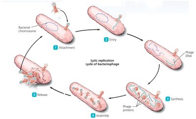
Bacteriophage Life Cycles: Lytic vs Lysogenic
Bacteriophages (viruses that infect bacteria) can follow two distinct life cycles: lytic and lysogenic. These cycles determine how the phage interacts with its host and the outcome of infection.
Lytic Cycle: The phage invades the host, replicates, and lyses the cell to release new virions. This results in rapid destruction of the host cell.
Lysogenic Cycle: The phage genome integrates into the host genome, becoming a prophage. The prophage is replicated as the host divides. Environmental signals can induce the prophage to enter the lytic cycle.
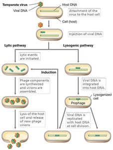
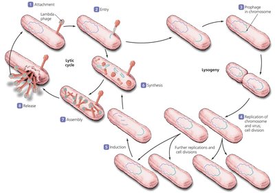
Connection Between Lytic and Lysogenic Cycles
The lysogenic cycle allows the phage genome to persist in the host without immediate destruction. Induction can trigger the switch to the lytic cycle, leading to active replication and cell lysis.

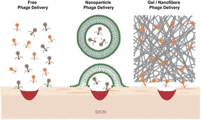
Bacteriophages as Treatments for Bacterial Infections
Lytic phages are being explored as alternatives to antibiotics for treating bacterial infections. They can be applied topically, orally, or via inhalation, and may be genetically modified for increased specificity.
Lytic phages: Destroy bacterial cells rapidly, making them ideal for infection control.
Temperate phages: Not preferred for treatment as they do not immediately lyse cells.
Animal Viruses: Types of Infections and Entry Mechanisms
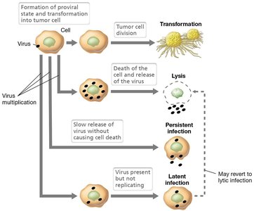
Types of Viral Infections in Animal Cells
Animal viruses can cause four main types of infections, each with distinct outcomes for the host cell:
Virulent/Lytic: Host cell is lysed to release virions.
Persistent: Host cell slowly releases virions without lysis.
Latent: Virus incorporates into host genome and remains dormant.
Transformation: Virus induces cancerous changes in host cells.

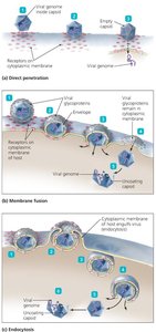
Animal Virus Entry and Uncoating
Animal viruses use specialized proteins for attachment and entry. There are three main entry mechanisms:
Direct Penetration: Capsid binds to membrane, genome enters cell, capsid remains outside.
Membrane Fusion: Envelope fuses with cell membrane, nucleocapsid enters cell.
Endocytosis: Entire virion is engulfed, nucleocapsid released from vesicle.
Uncoating is essential for viral genome accessibility and replication.
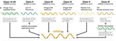
Viral Enzymes in Animal Viruses
Some animal viruses require unique enzymes for replication:
RNA-dependent DNA polymerase (Reverse Transcriptase): Converts RNA to DNA in retroviruses.
RNA-dependent RNA polymerase: Synthesizes RNA from RNA templates.
Host DNA/RNA polymerases: Used by DNA viruses for replication and transcription.
These enzymes are often packaged in the virion to ensure successful infection.

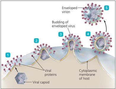
Viral Budding and Virion Formation
Enveloped viruses acquire their envelope from host membranes during budding. This process allows virions to exit the cell without lysis.
Budding: Nucleocapsid is wrapped in lipid membrane as it leaves the cell.
Protein-mediated cleavage: Some viruses require enzymes (e.g., neuraminidase in influenza) for release.
Laboratory Methods: Plaque Assays
Plaque Assays for Virus Quantification
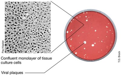
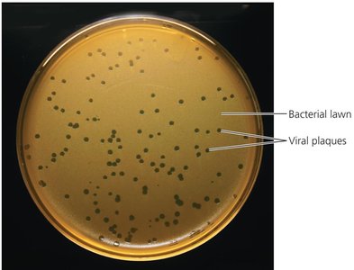
Plaque assays are used to measure viral titers in both bacteriophages and animal viruses. Plaques are cleared zones indicating cell lysis.
Bacterial lawn: Used for phage assays.
Monolayer of eukaryotic cells: Used for animal virus assays.
Plaque-forming units (PFU)/mL: Quantifies viral particles.
Viroids: Structure and Pathogenesis
Viroid Structure and Replication
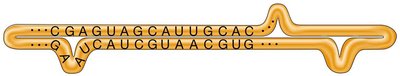
Viroids are infectious RNA molecules lacking a protein coat. They do not encode proteins and rely on host machinery for replication.
Small, circular, single-stranded RNA: 239-399 nucleotides.
Secondary structure: RNA folds to form double-stranded regions.
Replication: Uses host cell enzymes; cannot replicate independently.
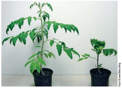
Viroid-Mediated Pathogenesis
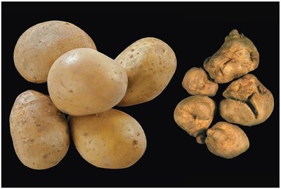
Viroids cause disease in plants, leading to growth defects, leaf and fruit malformation, and death. They alter gene expression by degrading specific plant mRNAs.
Prions: Structure, Pathogenesis, and Destruction
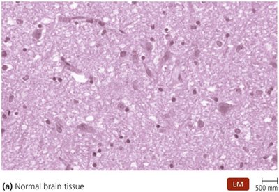
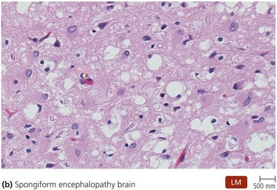
Prion Structure and Disease
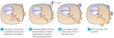
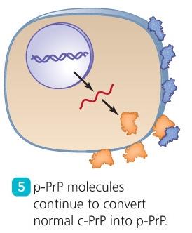
Prions are infectious proteins that cause transmissible spongiform encephalopathies in animals and humans. They lack a genome and propagate by inducing misfolding of normal proteins.
Diseases: Creutzfeldt-Jacob disease, kuru, mad cow disease, chronic wasting disease.
Spread: Depends on amino acid sequence compatibility between species.

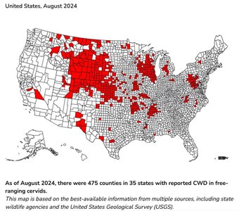
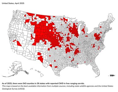
Chronic Wasting Disease (CWD) in Cervids
CWD is a prion disease affecting deer, elk, and related species. Prion proteins are found in various tissues and can be transmitted through bodily fluids.
Prion Destruction and Resistance
Prion aggregates are highly resistant to conventional sterilization methods. Extreme measures are required for decontamination.
Resistant to: Proteases, disinfectants, acid, boiling, radiation, autoclaving.
Recommended treatment: Immersion in 1N NaOH and autoclaving, or heating to 482˚C for 4 hours.
Enzymatic treatment: Approved in the EU for prion breakdown.
Comparison of Bacteria, Viruses, Viroids, and Prions
Structural and Functional Differences
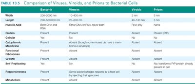
The following table summarizes the main differences between bacteria, viruses, viroids, and prions:
Feature | Bacteria | Viruses | Viroids | Prions |
|---|---|---|---|---|
Width | 200–1000 nm | 10–400 nm | 2–10 nm | 5 nm |
Nucleic Acid | Both DNA and RNA | Either DNA or RNA, never both | RNA | None (PrP protein) |
Protein | Present | Present | Absent | Present (PrP) |
Cytoplasmic Membrane | Present | Absent (some viruses have a membrane envelope) | Absent | Absent |
Functional Ribosomes | Present | Absent | Absent | Absent |
Growth | Present | Absent | Absent | Absent |
Self-Replicating | Present | Some bacteriophages respond to a host cell by injecting their genomes | Absent | No; transforms PrP protein already present in cell |
Metabolism | Present | Absent | Absent | Absent |
Additional info: These notes cover material from Chapter 13 (Viruses, Viroids, and Prions) and provide foundational knowledge for understanding acellular infectious agents in microbiology.
