 Back
BackAcellular Infectious Agents: Viruses, Viroids, and Prions
Study Guide - Smart Notes
 Tailored notes based on your materials, expanded with key definitions, examples, and context.
Important Types of Acellular Infectious Agents: Viruses
General Properties of Viruses
Viruses are acellular infectious agents that are classified as microbes due to their small size and ability to cause disease. Unlike prokaryotic and eukaryotic cells, viruses have a simple structure and lack many features of living organisms.
Acellular: Viruses are not composed of cells and are considered non-living.
Structure: All viruses possess a protein coat (capsid) and a nucleic acid genome (either DNA or RNA).
No Ribosomes or ATP Production: Viruses cannot synthesize proteins or generate their own energy.
Obligate Intracellular Parasites: Viruses must infect a host cell to replicate.
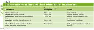
Comparison with Cellular Life: Viruses lack growth, metabolism, and cellular structure, distinguishing them from bacteria, archaea, and eukaryotes.
Characteristic | Bacteria, Archaea, Eukaryotes | Viruses |
|---|---|---|
Growth | Occurs in all | Does not occur |
Reproduction | Occurs in all | Host cell replicates the virus |
Responsiveness | Occurs in all | Reaction to host cells seen in some viruses |
Metabolism | Occurs in all | Uses host cell's metabolism |
Cellular structure | Present in all | Lacks cytoplasmic membrane or cellular structure |
Basic Structure of Viruses
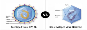
Capsid: Protein shell that encases the viral genome.
Nucleic Acid: Can be DNA or RNA, single- or double-stranded.
Envelope: Some viruses have a lipid envelope derived from the host cell membrane; others are non-enveloped (naked).
Shapes of Viruses
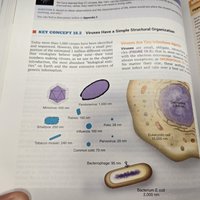
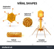
Viruses exhibit a variety of shapes, which are determined by the arrangement of their capsid proteins.
Polyhedral (Icosahedral): e.g., Adenovirus
Spherical: e.g., Influenza virus
Helical: e.g., Tobacco mosaic virus
Complex: e.g., Bacteriophage
Viral Host Range and Tropism
The host range of a virus refers to the spectrum of host species and cell types it can infect. Tropism describes the specific cells or tissues a virus targets within a host.
Broad Host Range: Some viruses (e.g., influenza, SARS-CoV-2) infect multiple species.
Narrow Host Range: Most bacteriophages infect only specific bacterial strains.
Tropism: Determined by viral surface proteins and host cell receptors (e.g., hepatitis viruses target liver cells; Ebola virus targets multiple organs).

Mechanisms of Viral Reproduction
Bacteriophage Reproduction
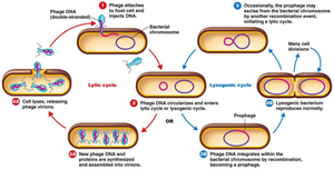
Bacteriophages (viruses that infect bacteria) can reproduce via two main cycles: the lytic cycle and the lysogenic cycle.
Lytic Cycle: Phage DNA is injected, host machinery is hijacked, new phages are produced, and the host cell is lysed.
Lysogenic Cycle: Phage DNA integrates into the host genome as a prophage and replicates with the host cell until induced to enter the lytic cycle.
Viral Reproduction in Animal Cells
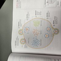
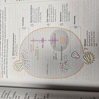
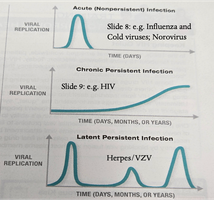
Animal viruses can cause acute, chronic, or latent infections. The process of viral entry and replication in animal cells differs from bacteriophages in several ways.
Acute Infections: Rapid onset, high viral production, and cell lysis (e.g., influenza, norovirus).
Chronic (Persistent) Infections: Virus persists in the host, often integrating into the genome (e.g., HIV forms a provirus).
Latent Infections: Virus remains dormant with periodic reactivation (e.g., herpes simplex virus, varicella zoster virus).
Key Differences: Entire virus enters animal cell; uncoating step releases genome; enveloped viruses often exit by budding rather than lysis.
Laboratory Cultivation of Viruses
Methods for Growing Viruses
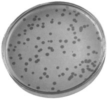
Bacteriophages: Grown on bacterial lawns in petri dishes, forming clear zones called plaques.
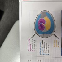
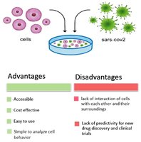
Animal Viruses: Grown in embryonated eggs, laboratory animals, or cell cultures.
Important Types of Acellular Infectious Agents: Viroids
Viroids
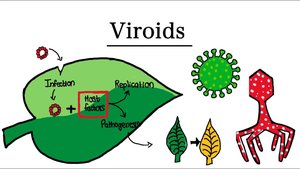
Viroids are infectious agents composed solely of a short strand of circular, single-stranded RNA. They lack a protein coat and are known to infect plants, causing various diseases.
Structure: Small, circular RNA molecules.
No Protein Coat: Distinguishes viroids from viruses.
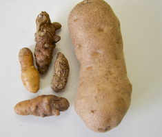
Pathogenicity: Cause diseases in plants, such as potato spindle tuber disease.
Important Types of Acellular Infectious Agents: Prions
Prions
Prions are infectious proteins that cause neurodegenerative diseases in humans and animals. They lack nucleic acids and propagate by inducing abnormal folding of normal cellular proteins in the brain.
Structure: Misfolded proteins (PrPSc).
Pathogenesis: Induce normal proteins to misfold, forming amyloid plaques that damage neural tissue.
Diseases: Creutzfeldt-Jakob disease, mad cow disease, scrapie, kuru.
Transmission: Can be acquired genetically, through contaminated food, medical procedures, or tissue transplants.
Diagnosis: Based on clinical symptoms, brain imaging, and detection of abnormal prion proteins.
Key Point: Prion diseases are always fatal and currently have no cure.
