 Back
BackAcellular Pathogens: Viruses, Viroids, and Prions – Structure, Classification, and Effects (chapter 6)
Study Guide - Smart Notes
 Tailored notes based on your materials, expanded with key definitions, examples, and context.
Nonviral Infectious Agents
Viroids, Virusoids, and Prions
Nonviral infectious agents include viroids, virusoids, and prions, each with unique structures and pathogenic mechanisms distinct from viruses.
Viroids: Short, circular RNA molecules capable of self-replication, lacking a protein coat. They are significant plant pathogens, causing diseases that impact agricultural crops.
Virusoids: Non-self-replicating single-stranded RNAs that require coinfection with a helper virus. Primarily plant pathogens, but some can infect animals. Satellite RNAs are a related group that also require a helper virus.
Prions: Infectious proteins that are misfolded forms of normal proteins. Prions can induce misfolding in normal proteins, leading to fatal neurodegenerative diseases (e.g., Transmissible Spongiform Encephalopathies). Prions are extremely resistant to standard sterilization techniques.
Example: Viroids cause diseases in plants such as the potato spindle tuber disease, leading to significant agricultural losses.

The Discovery of Viruses
Historical Milestones
The discovery of viruses marked a turning point in microbiology. Dmitri Ivanovski is credited with isolating the Tobacco Mosaic Virus (TMV) in 1892, demonstrating that infectious agents smaller than bacteria could cause disease. Martinus Beijerinck later coined the term "virus," meaning poison in Latin, after concluding that a filterable agent was responsible for the disease.

Viruses in the Biological Spectrum
General Properties and Evolution
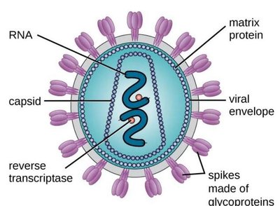
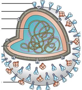
Viruses are the most abundant microbes on Earth and have played a role in the evolution of all domains of life. They are acellular (not cells) and are obligate intracellular parasites, meaning they require a living host cell for reproduction. The viral genome is composed of either DNA or RNA, never both, and is surrounded by a protein shell called a capsid. Viruses lack metabolic enzymes and protein synthesis machinery, relying entirely on the host cell for replication.
General Structure of Viruses
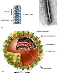
Capsids and Envelopes
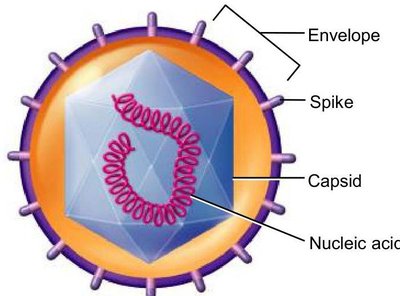
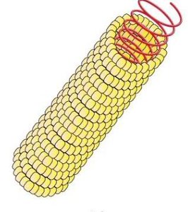
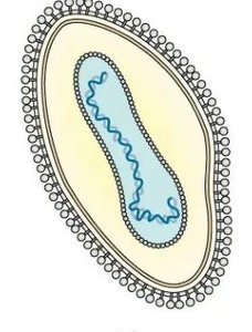
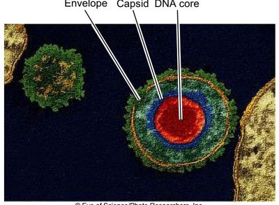
All viruses possess a capsid, a protein coat that protects the viral nucleic acid. The capsid is made of protein subunits called capsomers. The combination of the capsid and nucleic acid is termed the nucleocapsid. Some viruses have an additional external covering called an envelope, while those without are called naked viruses.
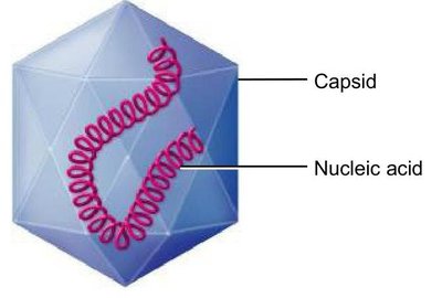
Capsid Types
Viruses exhibit three main capsid types:
Helical: Capsomers form a continuous helix, creating a cylindrical nucleocapsid.
Icosahedral: A 20-sided structure with 12 corners.
Complex: Atypical structures that do not fit the helical or icosahedral classification.

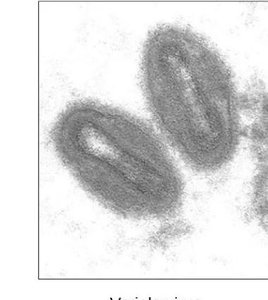
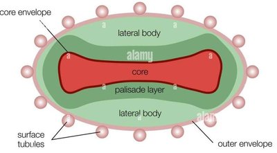
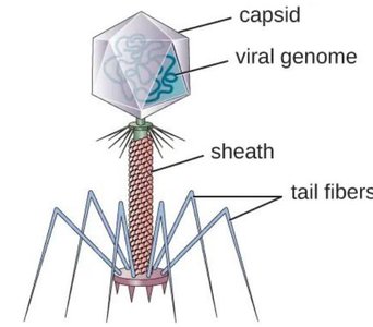
Complex Viruses
Some viruses, such as poxviruses and certain bacteriophages, have complex structures. Poxviruses lack a typical capsid and are covered by a dense layer of lipoproteins. Bacteriophages may have a polyhedral nucleocapsid with a helical tail and attachment fibers.
Functions of Capsid and Envelope
Protection and Attachment
The capsid protects the viral nucleic acid from degradation outside the host cell. Both enveloped and naked viruses have glycoprotein spikes that are crucial for recognizing and attaching to specific host cell receptors.
General Structure of Viral Envelope
Envelope and Spikes
The viral envelope is typically found in animal viruses and is acquired when the virus exits the host cell. Spikes are exposed proteins on the envelope's surface that are essential for attachment to the host cell.
Influenza Virus
Virulence Factors and Antigenic Variation
The influenza virus is characterized by glycoprotein spikes:
Hemagglutinin (H): Binds to host cells; multiple subtypes exist.
Neuraminidase (N): Hydrolyzes mucus and aids in viral budding and release; multiple subtypes exist.
Antigenic drift (small genetic changes) and antigenic shift (major genetic reassortment) in these proteins reduce immune effectiveness and can lead to pandemics.
Types of Viruses
Structural Classification
Viruses are classified based on their structure (helical, icosahedral, complex), presence or absence of an envelope, and the nature of their genome (DNA or RNA).

Viral Genome
Genetic Material
The viral genome contains the genes necessary for host invasion and replication. DNA viruses are usually double-stranded, while RNA viruses are typically single-stranded. Some RNA viruses have segmented genomes.
Classification of Viruses
Taxonomy and Nomenclature
Viruses are classified by structure, chemical composition, and genetic makeup. They are also grouped by host or tissue specificity. Family names end in -viridae, genus names in -virus, and species names are often binomial.
Persistent Infections
Latent and Chronic Infections
Persistent infections occur when a virus is not completely cleared and remains in the host:
Latent infection: Virus remains dormant and can reactivate (e.g., Varicella-zoster virus).
Chronic infection: Symptoms persist or recur over a long period (e.g., HIV).
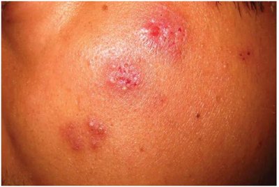
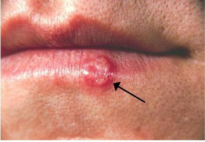
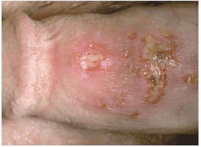
Herpes Simplex Virus (HSV)
Oral and Genital Herpes
HSV-1 typically causes oral herpes (cold sores), while HSV-2 is associated with genital lesions. Both are enveloped DNA viruses capable of latency and recurrent infections. Transmission occurs via direct contact, and antiviral medications can reduce symptoms and transmission risk.
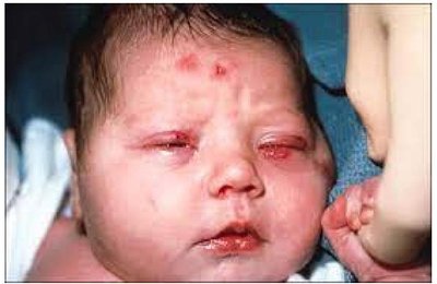
Neonatal Herpes
Transmission and Outcomes
Neonatal herpes, caused by HSV-1 or HSV-2, can be fatal in infants. Transmission can occur before or during birth. Outcomes include skin, eye, mouth disease, CNS disease, or disseminated disease. Early antiviral treatment improves prognosis.
Damage to Host Cell (Cytopathic Effects)
Cytopathic Effects (CPEs)
CPEs are observable cell abnormalities caused by viral infection, including:
Changes in cell size and shape
Inclusion body formation
Cell fusion (syncytium)
Cell lysis
Nuclear shrinkage
Transformation into cancerous cells (oncovirus)
Multiplication Cycle in Bacteriophages
Lytic and Lysogenic Cycles
Bacteriophages are viruses that infect bacteria. The lytic cycle results in host cell lysis and virus release, while the lysogenic cycle involves integration of the phage genome into the host chromosome as a prophage, which can later be induced to enter the lytic cycle.
Lytic cycle steps: Attachment, penetration, biosynthesis, maturation, lysis & release.
Lysogenic cycle: Integration, replication with host, induction, lytic cycle initiation.
Animal Virus Multiplication Cycle
General Phases
The multiplication of animal viruses involves attachment, penetration (endocytosis or fusion), uncoating, biosynthesis, assembly, and release (budding or lysis).
Attachment and Host Range
Host Specificity
Viruses attach to specific receptors on host cells, determining their host range (tropism). For example, poliovirus infects only primate intestinal and nerve cells, while rabies virus can infect many mammalian species.
Hepatitis Viruses
Types and Transmission
Hepatitis is liver inflammation caused by several unrelated viruses (HAV, HBV, HCV, HDV, HEV). Transmission varies: HAV and HEV are fecal-oral, while HBV, HCV, and HDV are bloodborne. Chronic infection increases the risk of liver cancer and cirrhosis.
Variation in Biosynthesis
Genome-Dependent Replication Strategies
Viral biosynthesis depends on genome type:
+ssRNA: Acts as mRNA, directly translated by host ribosomes.
-ssRNA: Must be transcribed into +ssRNA by viral RNA-dependent RNA polymerase before translation.
Retroviruses
Unique Replication Mechanism
Retroviruses are +ssRNA viruses that use reverse transcriptase to synthesize DNA from their RNA genome. This DNA is integrated into the host genome by integrase, forming a provirus. HIV is a well-known retrovirus.
Release of Viruses
Budding and Lysis
Enveloped viruses are released by budding (exocytosis), which does not immediately destroy the host cell. Naked and complex viruses are typically released by lysis, which ruptures and kills the host cell.
Cultivating and Identifying Viruses
Methods for Virus Growth
Viruses are obligate intracellular parasites and require living cells for replication. Common methods include:
Cell (tissue) cultures: Cultured cells support viral replication and allow observation of cytopathic effects.
Embryonated eggs: Used for growing certain viruses, especially for vaccine production.
Live animal inoculation: Occasionally used when other methods are unsuitable.
