 Back
Acid-Fast Stain: Principles, Procedure, and Clinical Significance
Study Guide - Smart Notes
 Tailored notes based on your materials, expanded with key definitions, examples, and context.
Acid-Fast Stain
Principles and Clinical Significance
The acid-fast stain is a differential staining technique used in microbiology to distinguish acid-fast bacteria, such as those in the genus Mycobacterium, from non-acid-fast bacteria. This method is especially important in clinical laboratories for identifying pathogens responsible for diseases like tuberculosis and leprosy. Acid-fast bacteria possess mycolic acids in their cell walls, which confer resistance to decolorization by acid-alcohol and impermeability to most aqueous stains.
Acid-fast bacteria: Retain the primary stain (carbolfuchsin) and appear fuchsia due to their waxy cell wall.
Non-acid-fast bacteria: Lose the primary stain during decolorization and are counterstained blue.
Clinical significance: Used to identify Mycobacterium tuberculosis, M. leprae, and other acid-fast organisms, including certain parasites.
Staining Procedure: Kinyoun Method
The Kinyoun method is a cold acid-fast staining procedure that uses a highly concentrated carbolfuchsin solution without heat. The steps are as follows:
Primary stain: Apply carbolfuchsin for 5 minutes to stain all cells fuchsia. Result: Both acid-fast and non-acid-fast cells appear fuchsia. Example: Mycobacterium tuberculosis (acid-fast) and E. coli (non-acid-fast) are used as controls.
Decolorization: Rinse with acid-alcohol for 10 seconds. Result: Acid-fast cells retain fuchsia color; non-acid-fast cells become colorless.
Counterstain: Apply methylene blue for 2 minutes. Result: Non-acid-fast cells are stained blue; acid-fast cells remain fuchsia.
Interpretation of Results
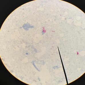
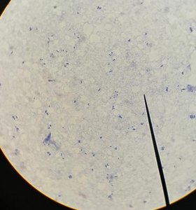
After staining, slides are observed under oil immersion. Acid-fast bacteria appear fuchsia, while non-acid-fast bacteria appear blue. This distinction is crucial for diagnosing diseases caused by acid-fast organisms.
Acid-fast reaction: Positive (fuchsia) for Mycobacterium tuberculosis; negative (blue) for E. coli.
Clinical application: Identification of tuberculosis, leprosy, and certain parasitic infections.
Comparison Table: Acid-Fast vs. Non-Acid-Fast Bacteria
Step | Acid-fast Bacteria | Non-Acid-fast Bacteria |
|---|---|---|
Carbolfuchsin | Fuchsia | Fuchsia |
Acid-alcohol | Fuchsia | Colorless |
Methylene blue | Fuchsia | Blue |
Clinical Relevance
Diseases caused by acid-fast bacilli (AFB):
Tuberculosis (Mycobacterium tuberculosis)
Leprosy (Mycobacterium leprae)
Acid-fast parasites: Example: Cryptosporidium causes cryptosporidiosis.
Microscopic Appearance
Acid-fast bacteria are observed as fuchsia-colored rods, while non-acid-fast bacteria appear blue under oil immersion microscopy.
Key Terms and Definitions
Acid-fast stain: A differential stain used to identify bacteria with waxy cell walls containing mycolic acids.
Carbolfuchsin: The primary stain in acid-fast procedures.
Acid-alcohol: The decolorizing agent.
Methylene blue: The counterstain for non-acid-fast bacteria.
Ziehl-Neelsen method: Traditional acid-fast staining method using heat.
Kinyoun method: Acid-fast staining method without heat, using concentrated carbolfuchsin.
Additional info:
The acid-fast stain is a critical diagnostic tool for identifying mycobacterial infections, especially in respiratory specimens.
Other differential stains include the Gram stain, which distinguishes between Gram-positive and Gram-negative bacteria based on cell wall structure.
