 Back
BackAcid-Fast Staining and Bacterial Growth: Microbiology Study Guide
Study Guide - Smart Notes
 Tailored notes based on your materials, expanded with key definitions, examples, and context.
Acid-Fast Staining
Principles and Importance
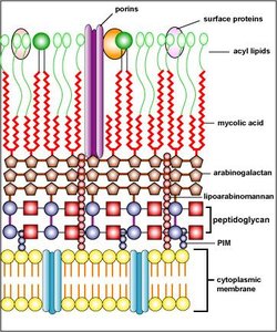
The acid-fast stain is a differential staining technique used to identify bacteria with unique cell wall properties, particularly those containing high levels of mycolic acid. This method is crucial for diagnosing diseases such as tuberculosis and leprosy, caused by Mycobacterium and Nocardia species. Acid-fast bacteria retain the primary stain (carbol fuchsin) even after treatment with acid-alcohol, due to their waxy, lipid-rich cell walls.
Key Feature: High concentration of mycolic acid in cell wall.
Clinical Relevance: Rapid detection of Mycobacterium tuberculosis and M. leprae.
Families: Mycobacteriaceae, Nocardiaceae, Gordoniaceae, Dietziacaea, Tsukamurellaceae.
Acid-Fast Staining Techniques
Two main methods are used for acid-fast staining: the Ziehl-Neelsen method and the Kinyoun method. Both utilize carbol fuchsin as the primary stain, but differ in their approach to dye penetration.
Ziehl-Neelsen Method: Uses heat to facilitate dye penetration.
Kinyoun Method: Increases phenol concentration in carbol fuchsin, acting as a chemical mordant and eliminating the need for heat.
Mordant: Substance that enhances dye fixation (heat or phenol).
General Steps:
Prepare a smear.
Add carbol fuchsin.
Apply acid-alcohol (decolorizer).
Add methylene blue (counterstain).
Mechanism of Acid-Fast Stain
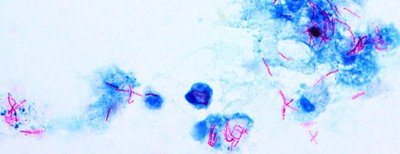
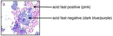
The acid-fast stain works due to the solubility of carbol fuchsin in phenol and its affinity for lipids. The dye penetrates the cell wall and is retained even after decolorization with acid-alcohol. Non-acid-fast bacteria lose the primary stain and take up the counterstain.
Acid-fast bacteria: Appear red/pink.
Non–acid-fast bacteria: Appear blue.
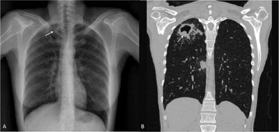
Clinical Application
Acid-fast staining is essential for identifying pathogens in clinical samples, especially in respiratory infections. It is used to detect acid-fast bacteria in lung tissue and monitor patients undergoing antibiotic therapy.
Comparison: Gram Stain vs. Acid-Fast Stain
The following table summarizes the differences between Gram stain and Acid-Fast stain:
Feature | Gram Stain | Acid-Fast Stain |
|---|---|---|
Purpose | Differentiates bacteria based on peptidoglycan structure | Identifies bacteria with mycolic acid in cell wall |
Primary Stain | Crystal violet (purple) | Carbol fuchsin (red) |
Mordant | Iodine | Heat (Ziehl–Neelsen) or phenol (Kinyoun) |
Decolorizer | Alcohol or acetone | Acid–alcohol |
Counterstain | Safranin (red/pink) | Methylene blue or brilliant green |
Result – Positive | Purple (Gram-positive) | Red/pink (acid-fast) |
Result – Negative | Pink/red (Gram-negative) | Blue/green (non–acid-fast) |
Key Cell Wall Feature | Peptidoglycan | Mycolic acids (waxy lipid) |
Clinical Use | General bacterial detection | Detection of Mycobacterium, Nocardia |
Turnaround | Minutes | Longer |
Limitations | Cannot detect acid-fast organisms well | Does not differentiate species |
Bacterial Growth
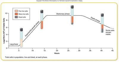
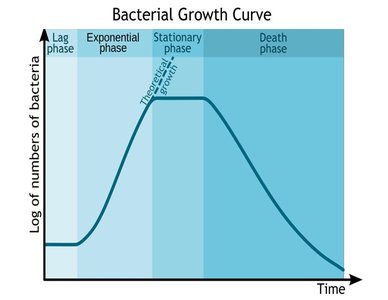
Bacterial Growth Curve
Bacterial populations grow in distinct phases, each characterized by specific physiological activities. Understanding these phases is essential for interpreting microbial behavior and the effects of antibiotics.
Lag Phase: Adaptation to environment; little or no cell division.
Exponential (Log) Phase: Rapid cell division; population increases exponentially.
Stationary Phase: Growth slows; new cells equal dying cells due to nutrient depletion and waste accumulation.
Death Phase: Decline in viable cells as resources are exhausted.
Effect of Antibiotics on Growth Curve
Antibiotics can alter the bacterial growth curve by affecting different phases:
Lag Phase: Extended if antibiotics delay growth initiation.
Exponential Phase: Reduced slope if antibiotics inhibit cell division.
Stationary Phase: May occur earlier if antibiotics affect metabolism or nutrient availability.
Death Phase: Accelerated by antibiotics inducing cell death.
Mathematical Models of Bacterial Growth
Bacterial growth can be described mathematically using exponential and doubling equations. These models assume ideal conditions without limiting factors.
Exponential Growth Equation:
N(t): Number of bacteria at time t
N_0: Initial number of bacteria
k: Growth rate constant
t: Time
e: Base of natural logarithm (≈2.718)
Example: Starting with 2,000 cells, k = 0.7/hr, after 5 hours:
Doubling Equation:
Example: Starting with 500 cells, generation time = 1 hour, after 4 hours:
Growth Rate Constant (K)
The growth rate constant (K) quantifies the number of generations per unit time during exponential growth.
Formula:
n: Number of generations
t: Number of hours
Unit: h-1
Possible Sources of Error in Wet Lab
Accurate staining and bacterial growth assessment require careful technique. Common errors include:
Using a loop that is too hot, killing bacteria.
Excessive heat during heat-fixing, damaging cells.
Leaving decolorizing agent on too long, removing too much stain.
Using old cultures, which may not stain properly.
Preparing smears that are too thick, obscuring results.
