 Back
BackAdaptive Immunity: Mechanisms and Cellular Players
Study Guide - Smart Notes
 Tailored notes based on your materials, expanded with key definitions, examples, and context.
Adaptive Immunity: The Third Line of Defense
Overview of Immune Defense Lines
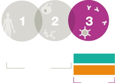
The immune system is organized into three main lines of defense: innate barrier defenses, innate cellular and molecular defenses, and adaptive defenses. The adaptive immune response is the body's third and final line of defense, providing a highly specific and long-lasting response to pathogens.
Innate Immunity: Immediate, non-specific defense mechanisms (e.g., skin, mucous membranes, phagocytes).
Adaptive Immunity: Delayed, highly specific response involving lymphocytes (T cells and B cells) and immunological memory.
Key Features of Adaptive Immunity
Specificity: Targets specific antigens unique to each pathogen.
Memory: Remembers previous encounters, resulting in a faster and stronger response upon re-exposure.
Delayed Response: Takes days to weeks to mount a primary response, but secondary responses are rapid and effective.
Interaction with Innate Immunity: Adaptive responses are activated when innate defenses are insufficient.
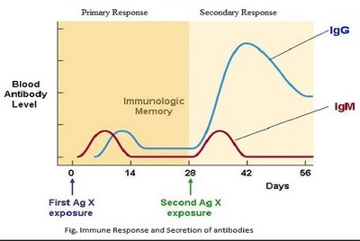
Example: Upon first exposure to an antigen, IgM antibodies are produced first, followed by IgG. Upon secondary exposure, IgG levels rise rapidly and to a higher level, demonstrating immunological memory.
Branches and Stages of Adaptive Immunity
Cellular and Humoral Responses
The adaptive immune system is divided into two main branches:
Cellular Response (T cell–mediated immunity): Involves T cells that target infected or abnormal cells.
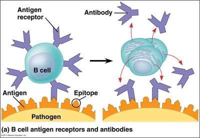
Humoral Response (B cell and antibody–mediated immunity): Involves B cells and the production of antibodies that neutralize pathogens in body fluids.
Both branches progress through four main stages: antigen presentation, lymphocyte activation, proliferation and differentiation, and antigen elimination with memory formation.
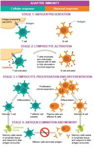
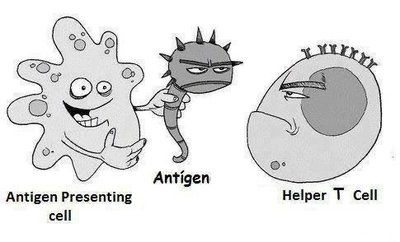
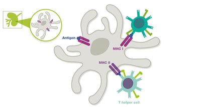
Stage 1: Antigen Presentation
Antigen-presenting cells (APCs), such as dendritic cells and macrophages, capture antigens and present them to T cells using major histocompatibility complex (MHC) molecules. B cells can recognize antigens directly without APCs.
APCs: Display processed antigens on their surface to T cells.
B Cells: Can bind directly to antigens via their B cell receptors (BCRs).
Stage 2: Lymphocyte Activation
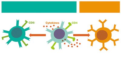
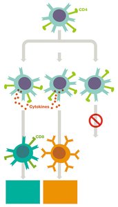
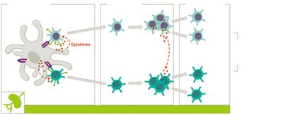
Upon antigen recognition, lymphocytes (T and B cells) are activated by cytokines. Activated T cells can further stimulate B cell activation, leading to a coordinated immune response.
Stage 3: Lymphocyte Proliferation and Differentiation
Activated lymphocytes undergo clonal expansion, producing many identical cells. These differentiate into:
Effector Cells: Actively engage in eliminating the antigen.
Memory Cells: Persist in lymphatic tissues for rapid response upon re-exposure.
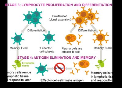
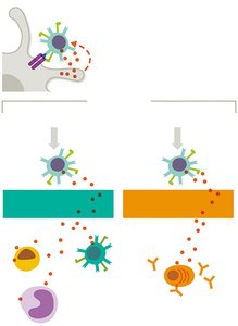
Stage 4: Antigen Elimination and Memory
Effector cells eliminate the antigen, after which most die off. Memory cells remain, providing long-term immunity.
Antigen Recognition and Immunogenicity
Antigen Structure and Immunogenicity
An antigen is any substance that can trigger an immune response. Most antigens are proteins or polysaccharides from pathogens, but they can also be lipids or small molecules (haptens).
Immunogenicity: The ability of an antigen to provoke an immune response. Proteins are the most immunogenic, followed by polysaccharides, then lipids.
Haptens: Small molecules that are only immunogenic when attached to a larger carrier molecule.
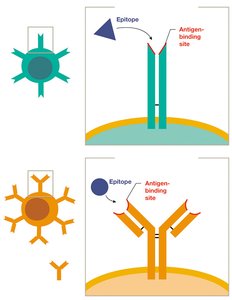
Epitopes and Antigen Receptors
Epitopes are specific regions on antigens recognized by B and T cells. Each lymphocyte expresses receptors (TCRs or BCRs) that recognize a single epitope.
Clonal Selection and Expansion
When a lymphocyte binds its specific epitope, it is activated and undergoes clonal expansion, producing effector and memory cells. B cells differentiate into plasma cells that secrete antibodies.
T Cells and B Cells: Types and Functions
T Cell Subtypes
T Helper Cells (TH, CD4+): Coordinate immune responses by releasing cytokines and activating other immune cells. Subclasses include TH1 (cellular response), TH2 (humoral response), and T regulatory cells (immune regulation).
T Cytotoxic Cells (TC, CD8+): Directly destroy infected, cancerous, or foreign cells.
B Cells
B Cells: Produce antibodies and present antigens. Upon activation, they differentiate into plasma cells (antibody-secreting) and memory B cells.
Summary Table: Comparing T Cells and B Cells
T cells | B cells | |
|---|---|---|
Adaptive immunity mediated | Cellular branch | Humoral branch |
Include | TH (CD4+), TC (CD8+) | Plasma cells |
Site of maturation | Thymus | Bone marrow |
Found in | Mainly lymphatic tissues | Mainly lymphatic tissues |
Antigen recognition receptors | TCRs | BCRs (antibodies) |
Require APC for activation? | Yes | No |
Memory cells made? | Yes | Yes |
MHC proteins present | MHC I | MHC I and II |
Considered APCs? | No | Yes |
Major Histocompatibility Complex (MHC) and Antigen Presentation
MHC I and MHC II Pathways
MHC I: Present on all nucleated cells; presents intracellular antigens to CD8+ T cytotoxic cells.
MHC II: Present only on APCs; presents extracellular antigens to CD4+ T helper cells.
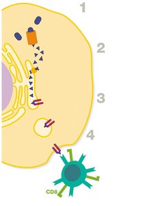
Example: Viral proteins produced inside an infected cell are processed and presented on MHC I molecules to cytotoxic T cells.
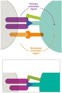
Activation of T Cells
T cells require two signals for full activation:
Primary Signal: TCR binds to MHC–antigen complex.
Secondary Signal: Co-stimulatory proteins (e.g., B7 on APC binds CD28 on T cell).
Clonal Expansion and Differentiation of T Cells
Activated T cells proliferate and differentiate into effector and memory cells. Cytokines released during this process determine the specific subclass of T helper cells (e.g., TH1, TH2).
Effector Functions and Immunological Memory
Antigen Elimination
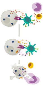
T Cytotoxic Cells: Release perforins and granzymes to induce apoptosis in infected or abnormal cells.
T Helper Cells: Release cytokines to activate B cells, cytotoxic T cells, and innate immune cells.

Immunological Memory
Memory T and B cells persist in lymphatic tissues, enabling a rapid and robust response upon subsequent exposures to the same antigen. This underlies the effectiveness of vaccines and long-term immunity.
Summary
Adaptive immunity is highly specific, involves memory, and is mediated by T and B lymphocytes.
It is divided into cellular (T cell-mediated) and humoral (B cell/antibody-mediated) branches.
Antigen presentation, lymphocyte activation, proliferation/differentiation, and antigen elimination/memory are the four main stages.
MHC molecules are essential for antigen presentation and self/non-self recognition.
Immunological memory ensures faster and stronger responses upon re-exposure to pathogens.
