 Back
BackAdaptive Immunity: Mechanisms and Features
Study Guide - Smart Notes
 Tailored notes based on your materials, expanded with key definitions, examples, and context.
Adaptive Immunity
Overview of Immune Defense Lines
The immune system is organized into three lines of defense: innate barrier defenses, innate cellular and molecular defenses, and adaptive defenses. The adaptive response is the body's third and final line of defense, providing a highly specific and effective mechanism for eliminating pathogens.
Innate Defenses: Immediate, non-specific responses including physical barriers and cellular mechanisms.
Adaptive Defenses: Custom-designed responses targeting specific antigens, involving cellular (T cell-mediated) and humoral (B cell-mediated) branches.
Characteristics of Adaptive Immunity
Adaptive immunity differs from innate immunity in several key ways. It takes longer to mount, is highly specific to particular antigens, and provides immunological memory for rapid and effective responses upon secondary exposure.
Specificity: Targets unique antigens.
Memory: Secondary exposure triggers a faster, more effective response, often preventing disease symptoms.
Immunocompromised: Individuals with impaired adaptive or innate systems are more susceptible to infections.
Branches and Stages of Adaptive Immunity
Cellular and Humoral Responses
The adaptive immune system is subdivided into two branches: cellular (T cell-mediated) and humoral (B cell and antibody-mediated) responses. Both branches aim to eliminate identified antigens and remember them for future encounters.
Cellular Response: T cells act in tissues and blood to destroy infected or abnormal cells.
Humoral Response: B cells and antibodies act in blood to neutralize pathogens.
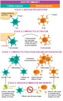
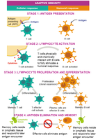
Four Stages of Adaptive Immune Response
Both cellular and humoral responses progress through four main stages:
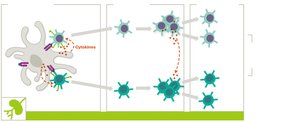
Antigen Presentation: Antigen-presenting cells (APCs) display antigens to T cells; B cells can directly interact with antigens.
Lymphocyte Activation: Lymphocytes are activated by cytokines after antigen presentation.
Lymphocyte Proliferation and Differentiation: Activated B and T cells undergo clonal expansion, forming effector and memory cells.
Antigen Elimination and Memory: Effector cells eliminate antigens; memory cells persist for rapid response upon re-exposure.
Key Cells and Antigen Recognition
T Cells and B Cells
T cells and B cells are the primary lymphocytes of adaptive immunity. T cells mature in the thymus, while B cells mature in the bone marrow. Both cell types reside mainly in lymphoid tissues and can recognize virtually any antigen due to gene shuffling mechanisms.
T Cells: Participate in both cellular and humoral immunity; mature in thymus.
B Cells: Coordinate humoral response by producing antibodies; mature in bone marrow.
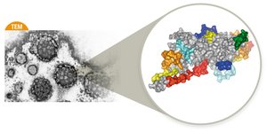
Antigen Features and Immunogenicity
Antigens are substances that trigger immune responses, typically proteins or polysaccharides from pathogens or abnormal cells. Immunogenicity depends on size, complexity, and composition, with proteins being most immunogenic.
Complete Antigens: Can trigger immune response independently.
Haptens: Incomplete antigens; require linkage to larger molecules for immunogenicity.

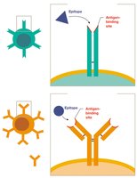
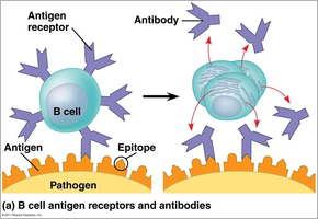
Epitopes and Antigen Recognition
Epitopes are specific regions of antigens recognized by B and T cells. Each lymphocyte has thousands of receptors, all recognizing the same epitope, but the body produces a vast array of lymphocytes for unlimited antigen recognition.
T Cell and B Cell Receptors
T cell receptors (TCRs) and B cell receptors (BCRs) are highly specific, recognizing only one epitope per lymphocyte. Antigen binding activates clonal expansion, producing effector and memory cells.
Effector Cells and Subtypes
Plasma Cells and Antibody Production
Activated B cells differentiate into plasma cells, which secrete antibodies (immunoglobulins) targeting the activating antigen.
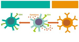
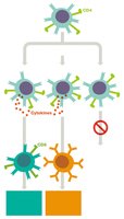
T Cell Subtypes
T cells differentiate into helper (TH) and cytotoxic (TC) cells, distinguished by CD proteins:
Helper T Cells (TH, CD4+): Coordinate immune responses by releasing cytokines and activating other leukocytes.
Cytotoxic T Cells (TC, CD8+): Directly destroy infected, cancerous, or foreign cells.
T Helper Cell Subclasses
Activated TH cells differentiate into subclasses with specialized functions:
TH1: Promote cellular response, activate TC cells, macrophages, and NK cells.
TH2: Promote humoral response, stimulate B cells.
Treg: Regulate immune response, suppress activity once threat is eliminated.
Self-Tolerance and MHC Screening
Screening for Self-Tolerance
T and B cells are screened for self-tolerance to prevent autoimmunity. T cells are screened in the thymus for recognition of self-MHC proteins, while B cells are screened in the bone marrow to ensure antibodies do not cross-react with self-antigens.
Stages of Adaptive Immunity: Detailed Mechanisms
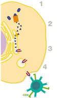
Stage 1: Antigen Presentation
APCs use MHC I or II to present antigens to T cells. MHC I is found on all nucleated cells and presents intracellular antigens to TC cells. MHC II is found only on APCs and presents extracellular antigens to TH cells.
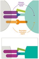
Stage 2: Lymphocyte Activation
T cells are activated in lymphatic tissues by APCs bearing MHC-antigen complexes. Activation requires two signals: TCR-MHC interaction and co-stimulatory protein binding (e.g., B7-CD28).
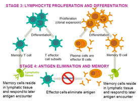
Stage 3: Lymphocyte Proliferation and Differentiation
Activated T and B cells undergo clonal expansion and differentiate into effector and memory cells. Cytokines influence the development of TH1 and TH2 cells, which direct cellular and humoral responses, respectively.
Stage 4: Antigen Elimination and Memory
Effector cells eliminate antigens, while memory cells persist in lymphatic tissues for rapid response upon re-exposure.
Humoral Response and Antibody Functions
B Cell Activation
B cells can be activated by T-dependent or T-independent antigens. T-dependent activation requires interaction with TH cells and co-stimulatory signals (CD40-CD40L).
Antibody Functions
Plasma cells secrete antibodies that bind to antigens, activating complement cascades, neutralizing antigens, and promoting phagocytosis.
Antibody Structure and Isotypes
Antibody Structure
Antibodies are composed of two heavy and two light chains, with antigen-binding sites at the tips. Each plasma cell produces antibodies recognizing the same epitope, but can switch isotypes.
Antibody Isotypes
IgG: Most abundant, found in all fluids, crosses placenta, activates complement, strong opsonin.
IgA: Prevalent in mucous membranes and secretions, neutralizes and opsonizes.
IgM: Early response, functions in agglutination and complement activation.
IgE: Low concentration, mediates allergic responses and fights parasites.
IgD: Sparsely represented, mainly on B cell surfaces, function unclear.
Immunological Memory and Adaptive Immunity Types
Immunological Memory
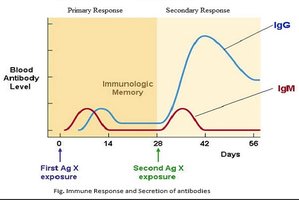
Memory cells allow for rapid, amplified responses upon re-exposure to antigens. Primary exposure generates IgM first, then IgG. Secondary exposure triggers a high titer of IgG and a small amount of IgM.
Types of Adaptive Immunity
Adaptive immunity can be acquired naturally or artificially, and is classified as active or passive:
Naturally Acquired Active Immunity: Infection triggers immune response; long-term protection.
Artificially Acquired Active Immunity: Vaccination triggers immune response; long-term protection.
Naturally Acquired Passive Immunity: Antibodies received non-medically (e.g., maternal); temporary protection.
Artificially Acquired Passive Immunity: Antibodies received medically (e.g., antiserum); temporary protection.
