 Back
BackAdaptive Immunity: Mechanisms, Cells, and Responses
Study Guide - Smart Notes
 Tailored notes based on your materials, expanded with key definitions, examples, and context.
Adaptive Immunity
Overview of Adaptive Immunity
Adaptive immunity is the body's highly specific defense mechanism against distinct pathogens and their products. It is characterized by the ability to recognize, respond to, and remember specific antigens, providing long-lasting protection.
Specificity: Targets unique antigens.
Inducibility: Activated in response to specific pathogens.
Clonality: Generates clones of lymphocytes specific to the antigen.
Unresponsiveness to self: Does not attack the body's own cells.
Memory: Mounts a faster and stronger response upon re-exposure to the same antigen.
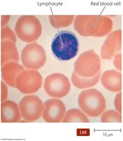
Adaptive immunity involves two main types of lymphocytes: B lymphocytes (B cells), which mature in the bone marrow, and T lymphocytes (T cells), which mature in the thymus. The two primary adaptive immune responses are cell-mediated and antibody-mediated (humoral) responses.
Elements of Adaptive Immunity: The Lymphatic System
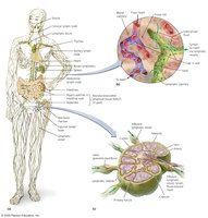
The lymphatic system is composed of lymphatic vessels, lymphoid cells, tissues, and organs. It screens the body's tissues for foreign molecules and is essential for the initiation and regulation of adaptive immune responses.
Lymphatic vessels: One-way system that returns lymph (fluid similar to plasma) from tissues to the circulatory system.
Primary lymphoid organs: Red bone marrow and thymus (sites of lymphocyte development).
Secondary lymphoid organs: Lymph nodes, spleen, tonsils, and mucosa-associated lymphoid tissue (MALT) (sites where immune responses are initiated).
Antigens and Their Properties
Definition and Types of Antigens
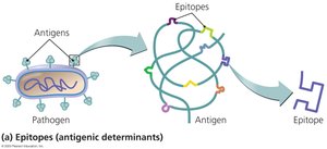
Antigens are molecules recognized as foreign by the immune system and capable of eliciting an immune response. They are identified by specific regions called epitopes or antigenic determinants.
Best antigens: Large, complex macromolecules (e.g., proteins from bacteria, viruses, fungi, protozoa).
Other sources: Food and dust particles can also contain antigens.
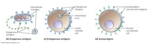
Types of Antigens
Exogenous antigens: Toxins and components of microbial cell walls, membranes, flagella, and pili.
Endogenous antigens: Produced by microbes that replicate inside host cells.
Autoantigens: Derived from normal cellular processes; usually not targeted by the immune system.
Major Histocompatibility Complex (MHC) and Antigen Presentation
Roles of MHC and Antigen-Presenting Cells
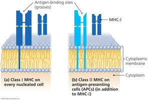
The major histocompatibility complex (MHC) consists of glycoproteins on cell membranes that present antigenic epitopes to immune cells. MHC molecules are crucial for distinguishing self from non-self and for initiating adaptive immune responses.
MHC class I: Present on all nucleated cells except red blood cells; present endogenous antigens.
MHC class II: Present only on antigen-presenting cells (APCs) such as macrophages, B cells, and dendritic cells; present exogenous antigens.
Immune Response Cytokines
Types and Functions of Cytokines
Cytokines are soluble regulatory proteins that act as intercellular signals in the immune system. They coordinate the activities of immune cells and are essential for the development and regulation of immune responses.
Interleukins (ILs): Signal among leukocytes.
Interferons (IFNs): Antiviral proteins that may act as cytokines.
Growth factors: Stimulate stem cell division.
Tumor necrosis factor (TNF): Kills tumor cells and regulates immune responses and inflammation.
Chemokines: Induce chemotaxis of leukocytes.
T Lymphocytes (T Cells)
Development and Function
T cells are produced in the red bone marrow and mature in the thymus. They circulate in the blood and lymph and migrate to secondary lymphoid organs. T cells are primarily responsible for cell-mediated immune responses, targeting cells infected with intracellular pathogens and abnormal cells (e.g., cancer cells).
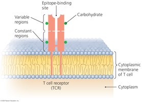
T Cell Receptor (TCR) Specificity
TCRs do not recognize free epitopes; they bind only to epitopes presented by MHC molecules on cell surfaces. This ensures that T cells act primarily against infected or abnormal cells.
Types of T Lymphocytes
Cytotoxic T lymphocytes (Tc): Directly kill infected or abnormal cells.
Helper T lymphocytes (Th): Regulate B cells and cytotoxic T cells; include Th1 and Th2 subsets.
Regulatory T lymphocytes (Treg): Suppress immune responses and promote tolerance to self-antigens.
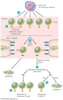
Clonal Deletion of T Cells
To prevent autoimmunity, T cells that react to self-antigens are eliminated through apoptosis during development in the thymus. Only T cells that recognize foreign antigens presented by MHC survive.
Cell-Mediated Immune Responses
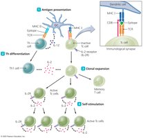
Activation of Cytotoxic T Cells
Cell-mediated responses are initiated in lymphoid organs and involve several steps:
Antigen presentation
Helper T cell differentiation
Clonal expansion
Self-stimulation
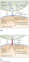
Mechanisms of Cytotoxic T Cell Killing
Perforin-granzyme pathway: Cytotoxic T cells release perforin and granzymes to induce apoptosis in target cells.
CD95 pathway: Involves interaction with CD95 (Fas) on target cells, triggering apoptosis.
Memory T Cells
Some activated T cells become memory T cells, which persist long-term in lymphoid tissues and respond rapidly upon re-exposure to their specific antigen.
B Lymphocytes (B Cells) and Antibodies
Development and Function
B cells mature in the bone marrow and are primarily found in the spleen, lymph nodes, and MALT. Their main function is the production and secretion of antibodies (immunoglobulins).
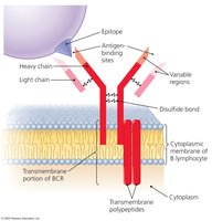
B Cell Receptor (BCR) Specificity
Each B cell expresses a unique BCR that binds a specific epitope. The diversity of BCRs allows the immune system to recognize millions of different antigens.
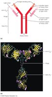
Antibody Structure and Function
Antibodies are Y-shaped proteins secreted by plasma cells (activated B cells). They have antigen-binding sites identical to the BCR of the parent B cell and mediate several immune functions:
Activation of complement and inflammation
Neutralization of toxins and pathogens
Opsonization (enhancing phagocytosis)
Agglutination (clumping of antigens)
Antibody-dependent cellular cytotoxicity (ADCC)
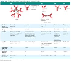
Classes of Antibodies
There are five main classes of antibodies, each with distinct functions and locations:
IgM: First antibody produced; effective in agglutination and complement activation.
IgG: Most abundant and long-lasting; crosses placenta; provides the majority of antibody-based immunity.
IgA: Found in body secretions (e.g., saliva, tears, breast milk); protects mucosal surfaces.
IgE: Involved in allergic responses and defense against parasitic infections.
IgD: Function not fully understood; found on B cell surfaces.
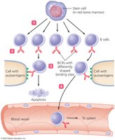
Clonal Deletion of B Cells
Self-reactive B cells are eliminated or inactivated in the bone marrow to prevent autoimmunity. Some may alter their BCRs instead of undergoing apoptosis.
Antibody Immune Responses
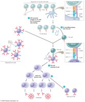
T-Dependent Antibody Immunity and Clonal Selection
Most antibody responses require the help of T helper cells (T-dependent). The process involves:
Antigen presentation for Th activation and proliferation
Differentiation of Th cells into Th2 cells
Activation of B cells
Proliferation and differentiation of B cells into plasma cells and memory cells
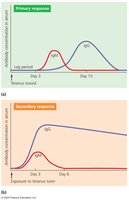
Immunological Memory
Memory B cells persist in lymphoid tissues and enable a rapid, robust secondary immune response upon re-exposure to the same antigen. The primary response is slower and produces fewer antibodies, while the secondary response is faster and stronger.
Types of Acquired Immunity
Active vs. Passive Immunity
Acquired immunity can be classified based on how it is obtained:
Naturally acquired active immunity: Response to antigens encountered in daily life (e.g., infection).
Naturally acquired passive immunity: Transfer of antibodies from mother to child (e.g., via placenta or breast milk).
Artificially acquired active immunity: Response to antigens introduced by vaccination.
Artificially acquired passive immunity: Administration of preformed antibodies (e.g., antiserum).
Type | Active | Passive |
|---|---|---|
Naturally Acquired | Exposure to pathogens in daily life | Maternal antibodies via placenta or milk |
Artificially Acquired | Vaccination | Injection of antibodies (antiserum) |
