 Back
BackLec 26:Adaptive Immunity: Mechanisms, Components, and Antibody Diversity
Study Guide - Smart Notes
 Tailored notes based on your materials, expanded with key definitions, examples, and context.
Adaptive Immunity: Overview
Definition and Main Functions
Adaptive immunity is a highly specific, learned defense system in vertebrates that targets particular pathogens (antigens), eliminates them, and retains memory for future protection. Unlike innate immunity, adaptive immunity utilizes B cells and T cells to generate tailored responses.
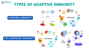
Antibody Production: B cells produce antibodies to neutralize antigens (humoral immunity).
Cellular Destruction: T cells kill infected cells and coordinate immune responses (cell-mediated immunity).
Immune Memory: The system responds faster and more effectively upon subsequent exposures to the same pathogen.
Key Components and Mechanisms
Lymphocyte Development
Lymphocytes (B and T cells) originate from stem cells in the bone marrow. These cells are central to adaptive immunity.
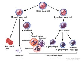
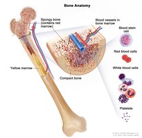
B Cells: Mature in the bone marrow and are responsible for antibody production in humoral immunity.
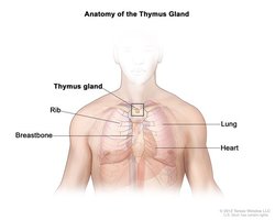
T Cells: Mature in the thymus and are responsible for cell-mediated immunity, including killing infected cells and coordinating immune responses.
Antigen Recognition
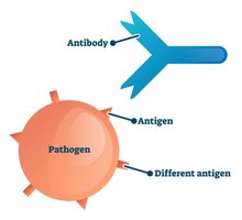
Adaptive immunity relies on the ability to distinguish self from non-self, targeting specific antigens present on pathogens.
Immune Memory
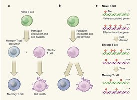
After an infection, some T and B cells differentiate into memory cells, which persist for years and provide rapid, robust protection upon re-exposure to the same pathogen.
Main Features of Adaptive Immunity
Specificity
Antibodies and cell receptors are precisely designed to match a specific antigen, functioning like a key in a lock.
Versatility
The adaptive immune system can respond to millions of unique invaders due to the diversity of antigen receptors.
Memory
Adaptive immunity retains memory of previous infections, enabling long-term protection.
Tolerance
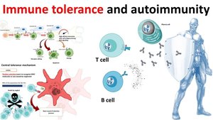
The system differentiates between self and non-self, preventing attacks on the body's own cells and maintaining immune tolerance.
The Adaptive Immune Response Process
Antigen Detection
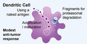
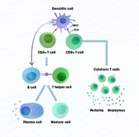
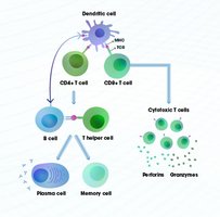
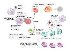
Dendritic cells capture antigens and present them to T cells in lymph nodes, initiating the adaptive response.
Activation
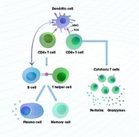
T cells are activated by antigen presentation and subsequently activate B cells.
Expansion and Differentiation
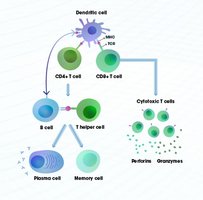
B cells proliferate and differentiate into effector cells (plasma cells) that produce antibodies.
Targeted Destruction
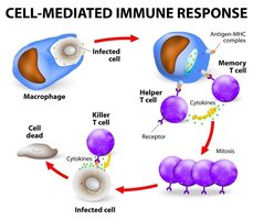
Antibodies and T cells work together to destroy pathogens.
Memory Formation
Long-lived memory B and T cells remain in the body for future protection.
Antibody Structure and Synthesis
Antibody Structure
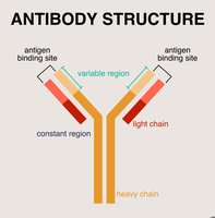
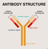
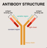
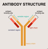
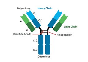
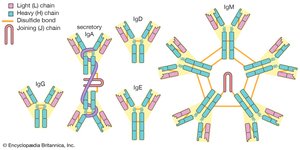
Antibodies (immunoglobulins) are large, Y-shaped proteins produced exclusively by B cells. They are composed of four polypeptide chains held together by disulfide bonds.
Heavy Chains: Two identical large chains.
Light Chains: Two identical smaller chains.
Variable (V) Region: Located at the tips of the Y arms, these regions form the antigen-binding site (paratope).
Constant (C) Region: The stem and lower arms, relatively uniform, determines antibody class and interaction with immune cells.
Hinge Region: Flexible segment allowing movement and binding to antigens at varying distances.
Synthesis and Secretion
Antibody synthesis involves activation, differentiation, and production:
Activation: Naïve B cells encounter their specific antigen and receive signals from Helper T cells.
Differentiation: Activated B cells proliferate and mature into plasma cells, expanding their internal machinery for protein production.
Production: Plasma cells secrete up to 2,000 antibody molecules per second.
Class Switching: B cells initially produce IgM, then switch to other classes (IgG, IgA) depending on infection type, while retaining the same antigen specificity.
Classes of Antibodies (Isotypes)
Overview
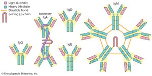
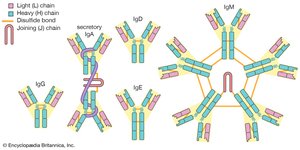
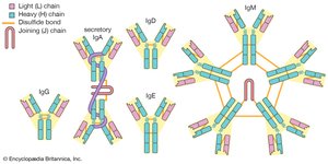
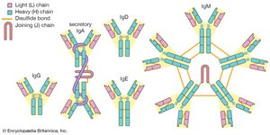
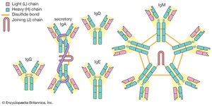
Antibodies are divided into five classes based on the structure of their heavy chain constant regions. These differences determine their location and function in the body.
Class | Structure | Main Function |
|---|---|---|
IgG | Monomer | Opsonization, neutralization, secondary response |
IgM | Pentamer | Agglutination, complement activation, primary response |
IgA | Dimer | Mucosal defense, colostrum protection |
IgE | Monomer | Allergy, binds mast cells/basophils |
IgD | Monomer | B cell receptor, rarely secreted |
IgG (Immunoglobulin G)
Most common antibody in circulation; effective at opsonization, neutralizing toxins/viruses, and primary antibody in secondary response.
IgM (Immunoglobulin M)
Largest antibody; pentameric structure with 10 binding sites, efficient at agglutination and complement activation.
IgA (Immunoglobulin A)
Most produced daily; guards mucosal surfaces, found in colostrum, often exists as a dimer.
IgE (Immunoglobulin E)
Lowest concentration in blood; binds mast cells/basophils, triggers histamine release in allergies.
IgD (Immunoglobulin D)
Rarely secreted; acts as a signaling sensor on naïve B cells.
Antibody Response Timeline
Primary vs. Secondary Response
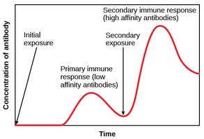
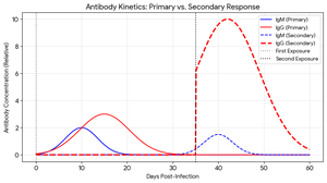
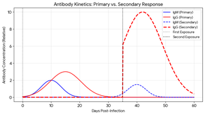
Upon first exposure to a pathogen, the adaptive immune system follows a predictable sequence of antibody production. The primary response is slower and less intense, while the secondary response is rapid and robust due to memory cells.
Lag Phase (Days 0–5): Innate system fights infection; adaptive system identifies threat.
IgM Rise (Days 5–10): IgM appears first, peaks around day 10–14, then declines.
IgG Rise (Days 10–21): IgG rises after IgM, peaks around week 3–4.
Convalescence: IgM disappears, IgG persists for long-term immunity.
T Cells: Linking Humoral and Cellular Immunity
Helper T Cells (CD4+)
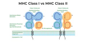
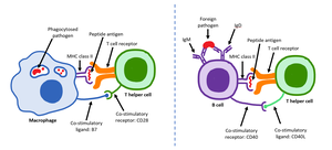
Helper T cells act as coordinators, linking cellular recognition to antibody production. They read antigen messages on MHC Class II molecules and activate B cells via cytokines.
Cytotoxic T Cells (CD8+)
Cytotoxic T cells scan for infected cells and induce apoptosis, handling cell-mediated immunity.
Major Histocompatibility Complex (MHC)
Role and Types
MHC molecules (HLA in humans) are surface glycoproteins that display peptide fragments to T cells, enabling the immune system to distinguish self from non-self.
MHC Restriction: T cells recognize antigens only when presented by self-MHC molecules.
Extreme Polymorphism: MHC genes are highly diverse, ensuring population-level protection.
Codominant Expression: Both parental MHC gene sets are expressed, increasing antigen presentation variety.
Cross-Presentation: Dendritic cells can present external antigens on MHC Class I to activate cytotoxic T cells.
Transplantation: Unique MHC molecules cause organ rejection.
Autoimmunity: Certain MHC alleles are linked to autoimmune diseases due to accidental presentation of self-peptides.
Summary Table: Adaptive Immunity Components
Component | Origin | Main Function |
|---|---|---|
B cell | Bone marrow | Antibody production (humoral immunity) |
T cell | Thymus | Cell-mediated immunity, coordination |
Antibody | B cell/plasma cell | Neutralization, opsonization, agglutination |
MHC | All nucleated cells | Antigen presentation |
Key Equations and Concepts
Antibody-Antigen Binding
The strength of antibody-antigen binding is termed affinity, and the overall effectiveness is avidity.
Clonal Expansion Formula
During immune response, lymphocytes undergo clonal expansion:
Where is the final number of cells, is the initial number, and is the number of divisions.
Antibody Concentration Over Time
Antibody concentration changes during primary and secondary responses:
Where is IgG concentration at time , is initial concentration, and is the decay constant.
Conclusion
Adaptive immunity is a sophisticated system that provides specific, versatile, and long-lasting protection against pathogens. Its core components—B cells, T cells, antibodies, and MHC molecules—work together to ensure targeted defense, immune memory, and tolerance. Understanding these mechanisms is fundamental for microbiology and immunology studies.
