Back
BackAdaptive Immunity: Specific Defenses of the Host
Study Guide - Smart Notes
 Tailored notes based on your materials, expanded with key definitions, examples, and context.
Adaptive Immunity: Specific Defenses of the Host
Introduction to Adaptive Immunity
Adaptive immunity is a specialized defense mechanism that targets specific pathogens after exposure. It is characterized by the ability to distinguish between "self" and "nonself" and is activated when innate defenses are insufficient. Adaptive immunity is acquired through infection or vaccination and involves two main responses:
Primary response: The first encounter with a particular foreign substance.
Secondary response: Subsequent encounters with the same substance, which are faster and more effective due to immunological memory.
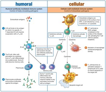
Dual Nature of the Adaptive Immune System
Humoral Immunity
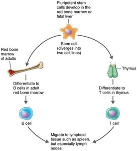
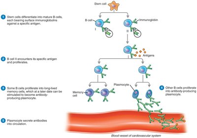
Humoral immunity involves the production of antibodies by B lymphocytes (B cells) that combat foreign molecules known as antigens. B cells are created and mature in the red bone marrow and, once mature, reside in the blood and lymphoid organs. They recognize antigens and produce antibodies.
Cellular Immunity (Cell-Mediated Immunity)
Cellular immunity is mediated by T lymphocytes (T cells), which recognize antigenic peptides processed by phagocytic cells. T cells mature in the thymus and reside in blood and lymphoid organs. T cell receptors (TCRs) on their surface contact antigens, leading to the secretion of cytokines rather than antibodies. Cellular immunity is most effective against virus-infected cells and intracellular bacteria.
Comparison of Humoral and Cellular Immunity
Cellular immunity: Attacks antigens that have already entered cells (e.g., viruses, intracellular bacteria).
Humoral immunity: Fights invaders and threats outside cells (e.g., extracellular bacteria, toxins, viruses before cell entry).
Cytokines: Chemical Messengers of Immune Cells
Cytokines are protein messengers produced in response to stimuli. They regulate the intensity and duration of immune responses by mediating communication between cells.
Interleukins (ILs): Communication between leukocytes.
Chemokines: Induce migration (chemotaxis) of leukocytes.
Interferons (IFNs): Interfere with viral infections of host cells.
Tumor necrosis factor alpha (TNF-α): Involved in inflammation of autoimmune diseases.
Hematopoietic cytokines: Control stem cell development into red and white blood cells.
Overproduction of cytokines can lead to a cytokine storm, which is harmful to the host.
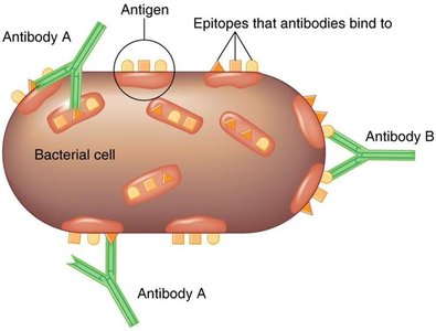
Antigens and Epitopes
Antigens are substances that provoke the production of antibodies, usually components of invading microbes or foreign substances (e.g., capsules, cell walls, flagella, fimbriae, toxins, viral capsids, and spikes). Nonmicrobial antigens include egg white, pollen, and cell surface molecules. Antibodies interact with specific regions on the antigen called epitopes (antigenic determinants).
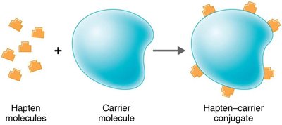
Haptens are molecules too small to be antigenic on their own; they must attach to carrier molecules to provoke an immune response.
Humoral Immunity: Antibodies (Immunoglobulins)
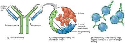
Antibodies are compact, soluble proteins called immunoglobulins (Ig) that recognize and bind to specific antigens, targeting them for destruction. The valence of an antibody is the number of antigen-binding sites it possesses (e.g., bivalent antibodies have two binding sites).
Each antibody consists of four protein chains forming a Y shape: two identical light chains and two identical heavy chains joined by disulfide bonds. The variable (V) regions at the ends of the arms bind epitopes, while the constant (Fc) region forms the stem and is identical for each Ig class. There are five classes of immunoglobulins: IgG, IgM, IgA, IgD, and IgE.
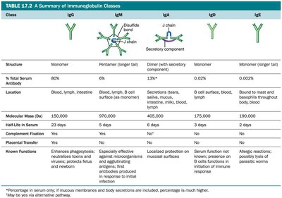
Classes of Immunoglobulins
Class | Structure | % Serum Antibody | Location | Functions |
|---|---|---|---|---|
IgG | Monomer | 80% | Blood, lymph, intestine | Crosses placenta, activates complement, enhances phagocytosis, neutralizes toxins/viruses |
IgM | Pentamer | 6% | Blood, lymph, B cell surface | First antibody produced, agglutination, activates complement |
IgA | Dimer (secretions), monomer (serum) | 13% | Secretions, blood, lymph | Protects mucosal surfaces |
IgD | Monomer | 0.02% | B cell surface, blood, lymph | Initiates immune response on B cells |
IgE | Monomer | 0.002% | Bound to mast cells, basophils, blood | Allergic reactions, lysis of parasitic worms |
Humoral Immunity Response Process
B cells reside in lymphoid organs and interact with antigens. Each B cell has over 100,000 membrane-bound immunoglobulins serving as antigen receptors. Clonal selection occurs when a B cell is activated by binding its specific antigen, leading to clonal expansion and differentiation into antibody-secreting plasmocytes and memory B cells.
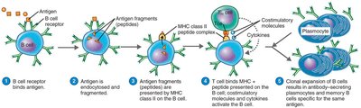
Activation of B Cells
T-dependent antigens: Require help from T helper cells (Th). Both B and T cells must recognize the antigen, and antigen presentation is required. The activated T cell produces cytokines that help activate the B cell.
T-independent antigens: Do not require T cell help. Many are polysaccharides from bacterial capsules or lipopolysaccharides (LPS).

Major Histocompatibility Complex (MHC)
MHC Class I: Present on all nucleated cells; presents antigens to cytotoxic T cells (Tc).
MHC Class II: Present on antigen-presenting cells (APCs) such as B cells, macrophages, and dendritic cells; presents antigens to T helper cells (Th).
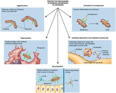
Results of Antigen–Antibody Interaction
When antibodies bind to antigens, they form an antigen–antibody complex. The strength of this bond is called affinity. These complexes protect the host by tagging foreign molecules or cells for destruction through several mechanisms:
Agglutination: Clumping of antigens.
Opsonization: Coating antigens to enhance phagocytosis.
Antibody-dependent cell-mediated cytotoxicity (ADCC): Target cell is lysed by immune cells.
Neutralization: Blocking attachment of toxins or viruses.
Activation of the complement system: Leads to cell lysis.
Cellular Immunity Response Process
T cells combat intracellular pathogens and abnormal host cells, such as cancer cells. They mature in the thymus, where thymic selection eliminates self-reactive T cells. T cells migrate to lymphoid tissues and attach to antigens via T-cell receptors (TCRs).
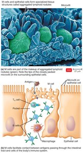
Pathogens entering the gastrointestinal tract pass through microfold cells (M cells) located over Peyer's patches, which transfer antigens to lymphocytes and APCs.
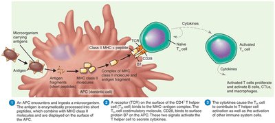
Antigen-Presenting Cells (APCs)
Dendritic cells (DCs): Engulf and degrade microbes, display antigens to T cells; found in skin, genital tract, lymph nodes, spleen, thymus, and blood.
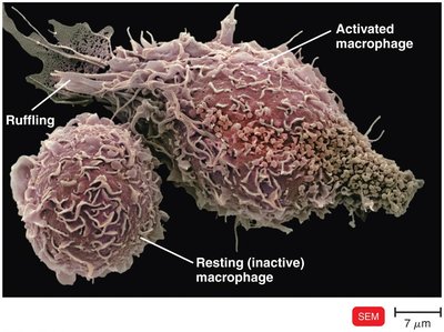
Macrophages: Activated by cytokines or antigen ingestion; migrate to lymph tissue to present antigens to T cells.
B cells: Also function as APCs.
Classes of T Cells
CD4+ (T helper cells, Th): Bind MHC class II molecules on APCs; produce cytokines to activate B cells and other immune cells.
CD8+ (Cytotoxic T lymphocytes, CTL): Bind MHC class I molecules; destroy infected or abnormal cells.
T Helper Cells (CD4+)
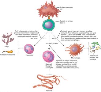
TCR on Th cells recognize and bind to antigen-MHC class II complexes on APCs. Costimulatory molecules activate Th cells, which then produce cytokines and differentiate into Th1, Th2, Th17, and memory cells.
Th1 cells: Produce IFN-γ, activate macrophages, and are involved in delayed hypersensitivity.
Th2 cells: Release IL-4, activate B cells to produce IgE, and activate eosinophils.
Th17 cells: Produce IL-17, contribute to inflammation, and recruit neutrophils.
T Regulatory Cells (Treg)
Treg cells are a subset of CD4+ cells that carry an additional CD25 molecule. They suppress T cells against self, protect intestinal bacteria required for digestion, and protect the fetus during pregnancy.
Cytotoxic T Lymphocytes (CD8+ CTLs)
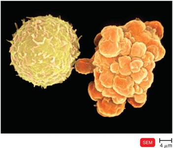
CTLs are activated when their TCR interacts with class I MHC and antigenic peptide on another body cell. Activated CTLs recognize and kill infected or abnormal cells by releasing perforin (forms pores) and granzymes (induce apoptosis).
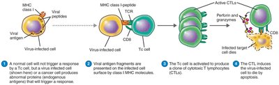
Apoptosis is programmed cell death, preventing the spread of infectious viruses. Cells undergoing apoptosis fragment their genome and form membrane blebs.
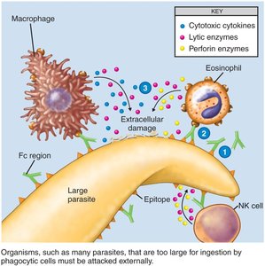
Nonspecific Cells and Extracellular Killing
Natural killer (NK) cells destroy virus-infected cells, tumor cells, and large extracellular parasites. They are not antigen-specific and detect target cells lacking MHC class I. NK cells induce apoptosis by forming pores in the target cell.
Antibody-Dependent Cell-Mediated Cytotoxicity (ADCC)
Protozoans and helminths, too large to be phagocytized, are coated with antibodies. Immune cells (NK cells, macrophages) attach to the Fc regions of antibodies and lyse the target cell by releasing cytotoxic chemicals.
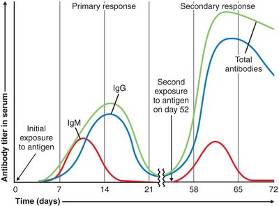
Immunological Memory
The primary response occurs upon first exposure to an antigen, while the secondary (memory) response is faster, more robust, and longer-lasting due to memory cells. Class switching from IgM to IgG, IgE, or IgA occurs during the secondary response. The antibody titer reflects the intensity of the humoral response.
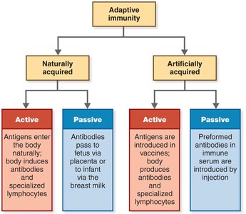
Types of Adaptive Immunity
Naturally acquired active immunity: Resulting from infection.
Naturally acquired passive immunity: Transfer of antibodies from mother to fetus or infant (transplacental or via colostrum).
Artificially acquired active immunity: Injection of vaccines (immunization).
Artificially acquired passive immunity: Injection of preformed antibodies.
Summary: Dual Nature of the Adaptive Immune System
The adaptive immune system consists of humoral (antibody-mediated) and cellular (cell-mediated) branches. Humoral immunity controls freely circulating pathogens, while cellular immunity controls intracellular pathogens. Both branches interact and coordinate to provide effective defense against a wide range of infectious agents.