 Back
(Chapter 17, Part 1) Adaptive Immunity: Specific Defenses of the Host
Study Guide - Smart Notes
 Tailored notes based on your materials, expanded with key definitions, examples, and context.
Adaptive Immunity: Specific Defenses of the Host
Introduction to Adaptive Immunity
Adaptive immunity is a highly specialized defense system that targets specific pathogens after exposure. It distinguishes 'self' from 'nonself' and is activated when innate defenses fail to stop a microbe. Adaptive immunity is acquired through infection or vaccination and involves two main responses:
Primary response: The first time the immune system combats a particular foreign substance.
Secondary response: Subsequent interactions with the same foreign substance; faster and more effective due to 'memory'.
The Dual Nature of the Adaptive Immune System
Humoral Immunity
Humoral immunity involves the production of antibodies that combat foreign molecules known as antigens. B cells are lymphocytes created and matured in red bone marrow, and they recognize antigens and make antibodies. In birds, B cells are named after the bursa of Fabricius, but in humans, they reside in the blood and lymphoid organs.
Cellular Immunity (Cell-Mediated Immunity)
Cellular immunity involves T lymphocytes, which recognize antigenic peptides processed by phagocytic cells. T cells mature in the thymus and reside in blood and lymphoid organs. T cell receptors (TCRs) on the T cell surface contact antigens, causing the T cells to secrete cytokines instead of antibodies. Cellular immunity is best at fighting virus-infected cells and intracellular bacteria.
Comparison of Humoral and Cellular Immunity
Cellular immunity attacks antigens that have already entered cells (e.g., viruses, some intracellular bacteria such as M. leprae and L. monocytogenes).
Humoral immunity fights invaders and threats outside cells (e.g., extracellular bacteria and toxins, viruses before they enter a host cell).
Cytokines: Chemical Messengers of Immune Cells
Cytokines are protein messengers produced in response to a stimulus. They include:
Interleukins (ILs): Communicate between leukocytes.
Chemokines: Induce migration of leukocytes.
Interferons (IFNs): Interfere with viral infections of host cells.
Tumor necrosis factor alpha (TNF-α): Involved in inflammation of autoimmune diseases.
Hematopoietic cytokines: Control stem cells that develop into red and white blood cells.
Overproduction of cytokines leads to a cytokine storm.
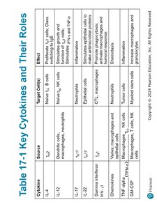
Key Cytokines and Their Roles
Type | Source | Target Cells | Function |
|---|---|---|---|
IL-1 | Macrophages | Helper T cells | Stimulates helper T cells |
IL-2 | Helper T cells | Helper T cells, cytotoxic T cells, NK cells | Stimulates proliferation |
IL-4 | Helper T cells | B cells | Promotes antibody production |
IL-17 | Helper T cells | Neutrophils | Promotes inflammation |
IFN-γ | T cells, NK cells | Macrophages | Activates macrophages |
TNF-α | Macrophages | Various cells | Promotes inflammation |
Additional info: | Other cytokines exist with specialized functions. |
Antigens and Their Properties
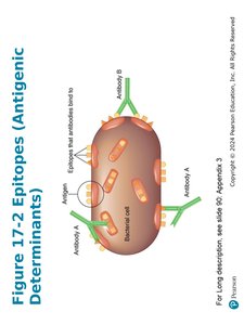
Antigens are substances that cause the production of antibodies. They are usually components of invading microbes or foreign substances, such as cell walls, flagella, fimbriae, toxins, capsules, and viral spikes. Nonmicrobial antigens may include egg white, pollen, and cell surface molecules.
Antibodies interact with specific regions (epitopes or antigenic determinants) on the antigen.
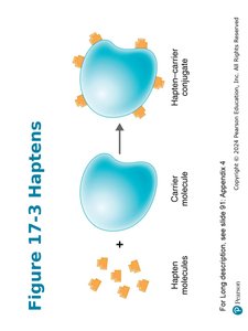
Haptens are molecules too small to be antigenic; they attach to carrier molecules and provoke an immune response.
Humoral Immunity: Antibodies
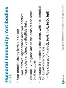
Antibodies, also known as immunoglobulins (Ig), are compact soluble proteins that recognize and bind to specific antigens, targeting them for destruction. The valence is the number of antigen-binding sites on an antibody; bivalent antibodies have two binding sites.
Four protein chains form a Y shape: two identical light chains and two identical heavy chains joined by disulfide links.
Variable (V) regions are at the ends of the arms and bind epitopes.
Constant (C) region is the stem, which is identical for a particular Ig class.
Five classes of Ig: IgG, IgM, IgA, IgD, IgE.
Immunoglobulin Classes
Class | Structure | Serum % | Location | Function |
|---|---|---|---|---|
IgG | Monomer | 80% | Blood, lymph, intestine | Crosses placenta, protects fetus, activates complement, enhances phagocytosis, neutralizes toxins/viruses |
IgM | Pentamer | 6% | Blood vessels | First response, agglutination, activates complement |
IgA | Monomer/dimer | 13% | Mucous membranes, secretions | Prevents microbial attachment |
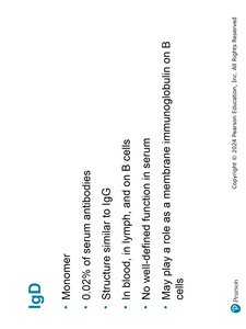
IgD | Monomer | 0.02% | Blood, lymph, B cells | Membrane immunoglobulin on B cells |
IgE | Monomer | 0.002% | Mast cells, basophils, blood | Histamine release, parasitic worms, allergies |
Humoral Immunity Response Process
B cells reside in and interact with antigens in lymphoid organs (spleen and lymph nodes). Each B cell has over 100,000 membrane-bound surface immunoglobulins that serve as antigen receptors. The process involves:
Clonal selection: B cell is activated when its B-cell receptor (BCR) binds to its antigen.
Clonal expansion: The activated B cell proliferates and differentiates into plasma cells (secrete antibody) and memory B cells.
Activation and Clonal Expansion of Antibody-Producing Cells
T-Dependent and T-Independent Antigens
T-dependent antigens: Require a T helper cell (Th) for activation; include proteins from viruses, bacteria, red blood cells.
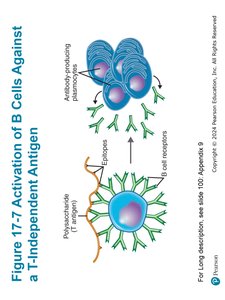
T-independent antigens: Do not need Th assistance; often polysaccharides from bacterial capsules or LPS.
Major Histocompatibility Complex (MHC)
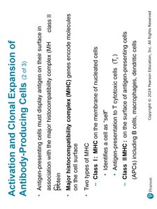
Antigen-presenting cells must display antigen on their surface in association with MHC class II molecules.
MHC genes encode molecules on the cell surface.
Class I MHC: On the membrane of nucleated cells; identifies cell as 'self'.
Class II MHC: On the surface of antigen-presenting cells (APCs), including B cells, macrophages, dendritic cells.
Activation Process
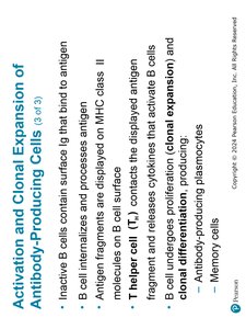
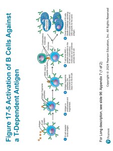
Inactive B cells contain surface Ig that bind to antigen.
B cell internalizes and processes antigen; antigen fragments are displayed on MHC Class II molecules on B cell surface.
T helper cell (Th) contacts the displayed antigen fragment and releases cytokines that activate B cells.
B cell undergoes proliferation (clonal expansion) and differentiation, producing antibody-producing plasma cells and memory cells.
The Diversity of Antibodies
The estimated extent of antibody diversity is 1011 (100 billion) different antibodies. Immunoglobulin genes have segments that can rearrange to produce diversity in the antigen-binding section of the antibody molecule. Clonal deletion removes potentially harmful self-reactive B cells.
Results of the Antigen-Antibody Interaction
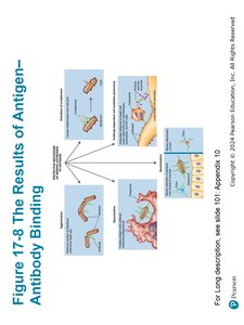
An antigen-antibody complex forms when antibodies bind to antigens. The strength of the bond is the affinity. Antibodies protect the host by tagging foreign molecules or cells for destruction. The main results include:
Agglutination
Opsonization
Antibody-dependent cell-mediated cytotoxicity
Neutralization
Activation of the complement system
Cellular Immunity Response Process
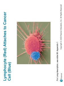
T cells combat intracellular pathogens and abnormal host cells such as cancer cells. They mature in the thymus, and thymic selection eliminates immature and self-reactive T cells. T cells migrate from the thymus to lymphoid tissues and attach to antigens via T-cell receptors (TCRs).
Pathogen Entry and M Cells
Pathogens entering the gastrointestinal tract pass through microfold cells (M cells) located over Peyer's patches (aggregated lymphoid nodules). These cells transfer antigens to lymphocytes and antigen-presenting cells (APCs).
Summary Table: Immunoglobulin Classes
Class | Structure | Serum % | Location | Function |
|---|---|---|---|---|
IgG | Monomer | 80% | Blood, lymph, intestine | Crosses placenta, protects fetus, activates complement, enhances phagocytosis, neutralizes toxins/viruses |
IgM | Pentamer | 6% | Blood vessels | First response, agglutination, activates complement |
IgA | Monomer/dimer | 13% | Mucous membranes, secretions | Prevents microbial attachment |
IgD | Monomer | 0.02% | Blood, lymph, B cells | Membrane immunoglobulin on B cells |
IgE | Monomer | 0.002% | Mast cells, basophils, blood | Histamine release, parasitic worms, allergies |
Additional info:
Adaptive immunity is a cornerstone of vaccine development and immunotherapy.
Memory cells are critical for long-term protection against pathogens.
Immunoglobulin diversity is generated by gene rearrangement and somatic hypermutation.
