 Back
BackAdaptive Immunity: Structure, Function, and Mechanisms
Study Guide - Smart Notes
 Tailored notes based on your materials, expanded with key definitions, examples, and context.
Adaptive Immunity
Innate and Adaptive Immunity: Comparison
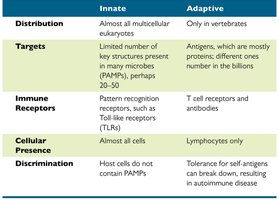
Immunity is divided into two main types: innate and adaptive. Innate immunity is present in almost all multicellular eukaryotes and provides a broad, non-specific defense. Adaptive immunity, found only in vertebrates, is highly specific and capable of remembering previous invaders.
Feature | Innate | Adaptive |
|---|---|---|
Distribution | Almost all multicellular eukaryotes | Only in vertebrates |
Targets | Limited number of key structures (PAMPs) | Antigens, mostly proteins; billions of types |
Immune Receptors | Pattern recognition receptors (e.g., TLRs) | T cell receptors and antibodies |
Cellular Presence | Almost all cells | Lymphocytes only |
Discrimination | Host cells do not contain PAMPs | Tolerance for self-antigens; breakdown can cause autoimmune disease |
Overview of Adaptive Immunity
Adaptive immunity is the body's ability to recognize and defend itself against distinct invaders and their products. It is characterized by specificity, inducibility, clonality, unresponsiveness to self, and memory.
Specificity: Targets specific antigens.
Inducibility: Activated in response to pathogens.
Clonality: Generates clones of immune cells.
Unresponsiveness to self: Prevents attack on self-antigens.
Memory: Remembers previous invaders for faster response.
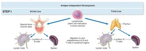
Lymphocytes and Types of Adaptive Immune Responses
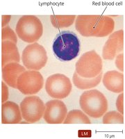
Adaptive immunity involves two main types of lymphocytes: B cells (mature in bone marrow) and T cells (mature in thymus). There are two types of adaptive immune responses: cell-mediated and antibody-mediated.
The Lymphatic System
Structure and Function
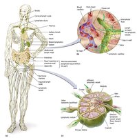
The lymphatic system screens tissues for foreign antigens and returns lymph to the circulatory system. Lymph is a fluid similar to blood plasma, arising from leaked blood vessel fluid.
Lymphoid Organs
Lymphoid organs are divided into primary (red bone marrow, thymus) and secondary (lymph nodes, spleen, tonsils, MALT) organs. Primary organs are sites of lymphocyte maturation; secondary organs are sites of immune response.

Antigens and Epitopes
Definition and Recognition
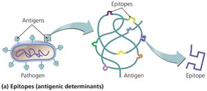
Antigens are molecules recognized as foreign and worthy of attack. Epitopes are three-dimensional regions on antigens recognized by immune cells. Antigens include bacterial components, viral proteins, and particles from food and dust.
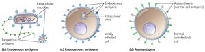
Types of Antigens
Antigens are classified as exogenous (external toxins and microbial components), endogenous (produced by microbes inside cells), and autoantigens (from normal cellular processes).
Self-Recognition and MHC
Major Histocompatibility Complex (MHC)
MHC is a group of glycoproteins found in cell membranes, important for tissue compatibility and antigen presentation. MHC molecules hold and position antigenic epitopes for immune cell recognition.

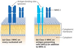
MHC Classes
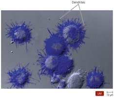
MHC class I proteins identify "self" and are present on all cells except red blood cells. MHC class II proteins present "non-self" antigens and are found on antigen-presenting cells (APCs) such as macrophages and dendritic cells.
Antigen Processing and Presentation
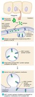
Mechanisms
Antigens must be processed for MHC proteins to display epitopes. Endogenous antigens are processed within infected cells, while exogenous antigens are processed by APCs.
T Lymphocytes (T Cells)
Development and Function
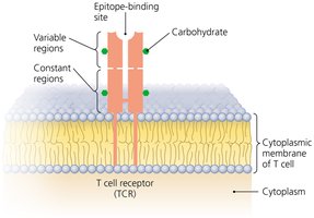
T cells are produced in the bone marrow and mature in the thymus. They circulate in lymph and blood, migrating to lymph nodes, spleen, and Peyer's patches. T cells have T cell receptors (TCRs) that bind epitopes associated with MHC proteins.
T Cell Receptor (TCR) Specificity
TCRs do not recognize epitopes directly; they only bind epitopes presented by MHC proteins. T cells act against cells harboring intracellular pathogens, cancer cells, and transplanted cells.
Types of T Lymphocytes
There are three main types of T cells based on surface glycoproteins and functions:
Cytotoxic T lymphocyte: Directly kills other cells.
Helper T lymphocyte: Regulates B cells and cytotoxic T cells; includes type 1 and type 2 helper T cells.
Regulatory T lymphocyte: Represses adaptive immune responses.
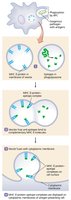
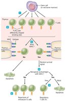
Clonal Deletion of T Cells
Clonal deletion ensures immune responses are not directed against autoantigens. Immature T cells undergo apoptosis if they do not recognize MHC or if they recognize autoantigens. Some self-recognizing T cells become regulatory T cells; others become protective T cells.
B Lymphocytes (B Cells) and Antibodies
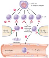
B Cell Function and Receptor Diversity
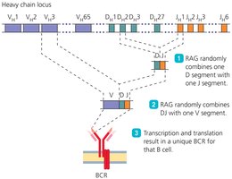
B cells secrete antibodies and each B cell makes a unique B Cell Receptor (BCR) that recognizes a single epitope. The diversity of BCRs allows recognition of millions of different epitopes. Diversity is generated by the RAG enzyme combining variable region gene segments.
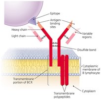
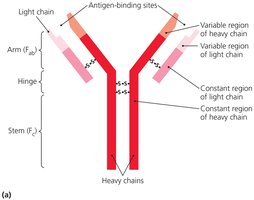
Antibody Structure
Antibodies are secreted by plasma cells and have antigen-binding sites identical to the BCR of the activated B cell. They consist of four peptide chains (two heavy, two light), two antigen-binding sites (Fab), flexible hinge regions, and a constant region (Fc).
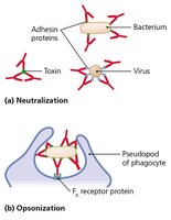
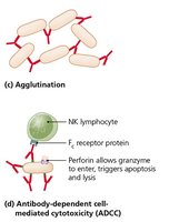
Functions of Antibodies
Antibodies perform several functions, including activation of complement and inflammation, neutralization, opsonization, agglutination, and antibody-dependent cellular cytotoxicity (ADCC).
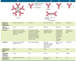
Classes of Antibodies
There are five classes of antibodies, each with distinct functions and locations:
IgM: First antibody produced.
IgG: Most common and longest-lasting antibody.
IgA: Associated with body secretions.
IgE: Involved in response to parasitic infections and allergies.
IgD: Function not well understood.
Class | Structure | Function | Location | Half-life |
|---|---|---|---|---|
IgM | Pentamer | Complement activation, agglutination | Serum | 5 days |
IgG | Monomer | Opsonization, neutralization | Serum, tissues | 23 days |
IgA | Dimer | Secretory immunity | Secretions | 6 days |
IgE | Monomer | Allergy, parasitic defense | Serum, tissues | 2 days |
IgD | Monomer | Unknown | B cell surface | 3 days |
Clonal Deletion of B Cells
Clonal deletion of B cells occurs in the bone marrow. Self-reactive B cells may become inactive or change their BCR rather than undergo apoptosis.
Immune Response Cytokines
Cytokine Types and Functions
Cytokines are soluble regulatory proteins acting as intercellular signals. Types include interleukins (ILs), interferons (IFNs), growth factors, tumor necrosis factor (TNF), and chemokines.
Interleukins (ILs): Signal among leukocytes.
Interferons (IFNs): Antiviral proteins, may act as cytokines.
Growth factors: Stimulate stem cell division.
Tumor necrosis factor (TNF): Kills tumor cells, regulates immune responses.
Chemokines: Signal leukocytes to move.
Cell-Mediated Immune Responses
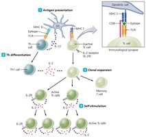
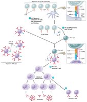
Activation of Cytotoxic T Cells
Cell-mediated responses target intracellular pathogens and abnormal cells. Steps in cytotoxic T cell activation include antigen presentation, helper T cell differentiation, clonal expansion, and self-stimulation.
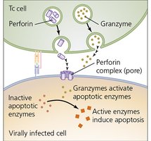
Mechanisms of Cytotoxic T Cell Killing
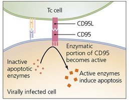
Cytotoxic T cells kill targets via two pathways:
Perforin-granzyme pathway: Synthesis of killing proteins induces apoptosis.
CD95 pathway: Mediated through glycoprotein interaction, also results in apoptosis.
Memory T Cells and Regulation
Some activated T cells become memory T cells, persisting for months or years and responding rapidly to subsequent antigen exposure. Regulatory T cells moderate cytotoxic T cell activity to prevent autoimmunity.
Antibody Immune Responses
T-Dependent Antibody Response
Antibody responses are mounted against exogenous pathogens and toxins. T-dependent responses require helper T cells and involve antigen presentation, helper T cell differentiation, B cell activation, and B cell proliferation.
Plasma Cells and Memory B Cells
Plasma cells secrete antibodies complementary to specific antigens and are short-lived. Memory B cells persist in lymphoid tissue and initiate rapid antibody production upon re-exposure to antigen.
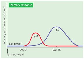
Immunological Memory
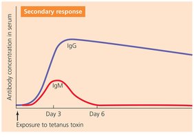
Primary immune response produces small amounts of IgM, taking days to eliminate antigen. Secondary response is faster and dominated by IgG, known as the anamnestic response.
Acquired Immunity Types
Four Types of Acquired Immunity
Naturally acquired active immunity: Exposure to antigen via infection.
Naturally acquired passive immunity: Maternal antibodies via placenta or colostrum.
Artificially acquired active immunity: Vaccination with antigens.
Artificially acquired passive immunity: Injection of antibodies (e.g., antivenom).
Additional info: Artificially acquired passive immunity provides rapid, short-term protection without immunological memory.
