 Back
BackAdaptive Immunity: Structure, Function, and Mechanisms
Study Guide - Smart Notes
 Tailored notes based on your materials, expanded with key definitions, examples, and context.
Overview of Adaptive Immunity
Attributes and Types of Adaptive Immunity
Adaptive immunity is the body's highly specific defense mechanism against distinct pathogens and their products. It is characterized by several unique attributes that distinguish it from innate immunity.
Specificity: Targets specific molecular shapes (epitopes) on antigens.
Inducibility: Immune cells are activated only in response to specific pathogens.
Clonality: Activated cells proliferate to form clones of identical cells.
Unresponsiveness to Self: Normally does not target the body's own cells.
Memory: Remembers previous encounters and responds more rapidly upon re-exposure.
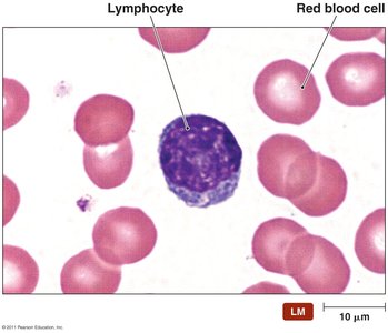
Adaptive immunity involves two main types of lymphocytes:
B lymphocytes (B cells): Mature in the bone marrow; responsible for humoral immunity.
T lymphocytes (T cells): Mature in the thymus; responsible for cell-mediated immunity.
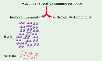
There are two main branches of adaptive immune responses:
Humoral immune responses: Mediated by B cells and antibodies, targeting extracellular pathogens.
Cell-mediated immune responses: Mediated by T cells, targeting intracellular pathogens.

Elements of Adaptive Immunity
The Lymphatic System
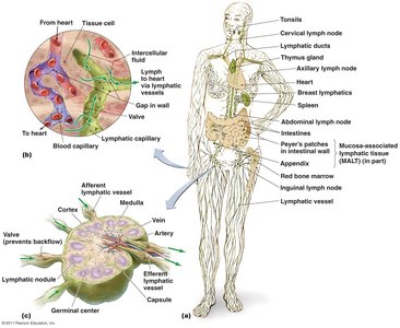
The lymphatic system screens the body's tissues for foreign antigens and is composed of lymphatic vessels, cells, tissues, and organs. It is essential for the initiation and regulation of adaptive immune responses.
Lymphatic vessels: One-way system that returns lymph (fluid similar to plasma) from tissues to the circulatory system.
Primary lymphoid organs: Red bone marrow and thymus (sites of lymphocyte development).
Secondary lymphoid organs: Lymph nodes, spleen, tonsils, and mucosa-associated lymphatic tissue (MALT).
Antigens and Epitopes
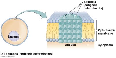
Antigens are molecules recognized as foreign and capable of provoking an immune response. They are identified by specific regions called epitopes (antigenic determinants).
Exogenous antigens: Originate outside host cells (e.g., bacteria, toxins, parasites).
Endogenous antigens: Generated within host cells (e.g., viral proteins).
Autoantigens: Self-molecules that can trigger autoimmunity if not properly regulated.
B Lymphocytes (B Cells) and Antibodies
B Cell Receptors (BCRs) and Antibody Structure
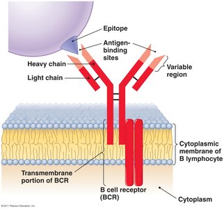
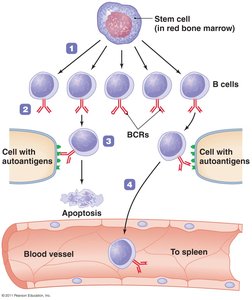
B cells arise and mature in the red bone marrow and are primarily found in the spleen, lymph nodes, and MALT. Their main function is the secretion of antibodies. Each B cell expresses a unique B cell receptor (BCR) that binds a specific epitope.
BCRs: Composed of two variable regions forming the antigen-binding sites; each B cell produces a single BCR specificity.
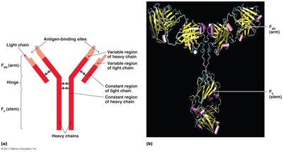
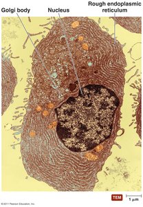
Antibodies (immunoglobulins): Secreted by plasma cells; structurally similar to BCRs with variable (Fab) and constant (Fc) regions.
Functions of Antibodies
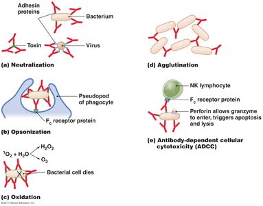
Antibodies contribute to immune defense through several mechanisms:
Neutralization: Block attachment of pathogens or toxins to host cells.
Opsonization: Tag pathogens for phagocytosis by immune cells.
Agglutination: Clump pathogens together for easier clearance.
Activation of complement: Trigger the complement cascade leading to pathogen lysis.
Antibody-dependent cellular cytotoxicity (ADCC): Recruit natural killer cells to destroy antibody-coated targets.
Classes of Antibodies
There are five main classes of antibodies, each with distinct roles:
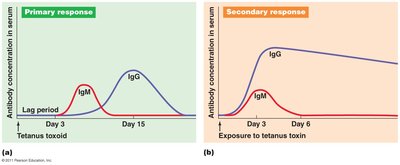
IgM: First antibody produced; effective in agglutination and complement activation.
IgG: Most abundant and long-lasting; crosses placenta, neutralizes toxins, and activates complement.
IgA: Found in body secretions; important for mucosal immunity.
IgE: Involved in allergic responses and defense against parasites.
IgD: Function not fully understood.
T Lymphocytes (T Cells)
T Cell Receptors (TCRs) and Types of T Cells
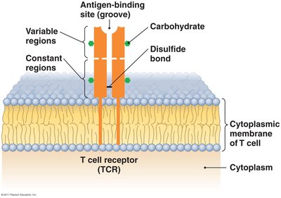
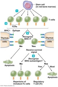
T cells are produced in the bone marrow and mature in the thymus. They circulate in the blood and lymph and are found in secondary lymphoid organs. TCRs recognize antigenic peptides only when presented by major histocompatibility complex (MHC) molecules.
Cytotoxic T lymphocytes (Tc): Kill infected or abnormal cells directly.
Helper T lymphocytes (Th): Regulate activities of B cells and cytotoxic T cells via cytokine secretion.
Regulatory T lymphocytes (Tr): Suppress immune responses to prevent autoimmunity.
Clonal Deletion and Self-Tolerance
To prevent autoimmunity, lymphocytes that react to self-antigens are eliminated through a process called clonal deletion. Cells that recognize autoantigens undergo apoptosis during development.
Immune Response Cytokines
Types and Functions of Cytokines
Cytokines are soluble regulatory proteins that mediate communication between immune cells. They regulate immune responses, inflammation, and cell growth.
Interleukins (ILs): Signal among leukocytes; over 35 types identified.
Interferons (IFNs): Inhibit viral replication and act as cytokines.
Growth factors: Stimulate stem cell division.
Tumor necrosis factor (TNF): Kills tumor cells and regulates immune responses.
Chemokines: Attract leukocytes to sites of infection.
Major Histocompatibility Complex (MHC) and Antigen Presentation
Classes of MHC Proteins
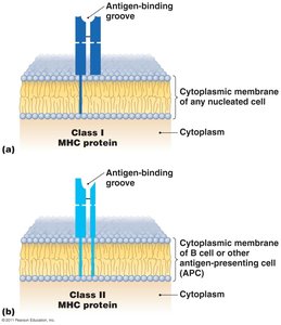
MHC molecules are glycoproteins on cell surfaces that present antigenic peptides to T cells. They are crucial for distinguishing self from non-self and for tissue compatibility in transplantation.
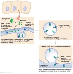
MHC Class I: Present on all nucleated cells; present endogenous antigens to cytotoxic T cells (CD8+).
MHC Class II: Present on antigen-presenting cells (APCs) such as dendritic cells, macrophages, and B cells; present exogenous antigens to helper T cells (CD4+).
Antigen Processing and Presentation
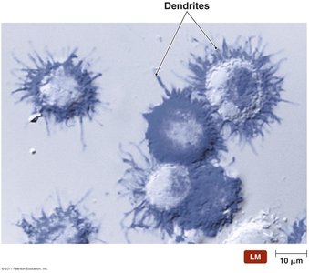
Antigen-presenting cells (APCs) process protein antigens and display peptide fragments on their surface bound to MHC molecules for recognition by T cells.
Dendritic cells: Activate naive T cells; found near skin and mucosa.
Macrophages: Present antigens and activate T cells.
B lymphocytes: Can also present antigens to T cells.
Cell-Mediated Immune Responses
Activation and Function of Cytotoxic T Cells
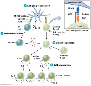
Cytotoxic T cells (Tc) are activated through a multi-step process involving antigen presentation, helper T cell differentiation, clonal expansion, and self-stimulation. Recognition of MHC is mediated by co-receptors CD4 (helper T cells) and CD8 (cytotoxic T cells).
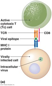
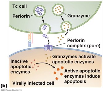
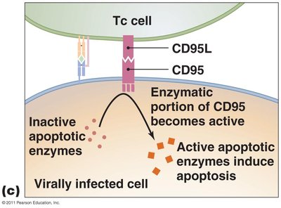
Mechanism: Tc cells recognize infected cells via MHC I-peptide complexes, bind through TCR and CD8, and induce apoptosis in the target cell using perforin, granzymes, and proteases.
Memory T Cells and Regulation
Some activated T cells become memory T cells, which persist long-term and respond rapidly upon re-exposure to their specific antigen. T cell responses are tightly regulated to prevent autoimmunity, requiring additional signals from APCs for full activation.
Humoral Immune Responses
Activation of B Cells and Antibody Production
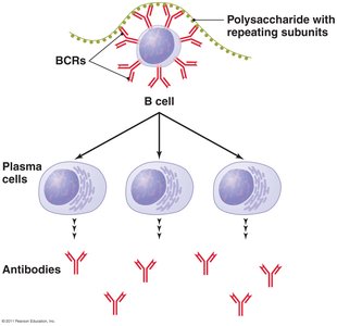
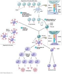
Humoral immunity targets exogenous pathogens and is mediated by B cells. B cells can be activated independently (T-independent antigens) or with T cell help (T-dependent antigens). Upon activation, B cells proliferate and differentiate into plasma cells that secrete antibodies.
Immunological Memory
Memory B cells are produced during B cell proliferation but do not secrete antibodies. They persist in lymphoid tissues and initiate rapid antibody production upon re-exposure to the same antigen, forming the basis of long-term immunity.
Types of Acquired Immunity
Active and Passive Immunity
Acquired immunity can be classified based on how it is obtained:
Naturally acquired: Response to antigens encountered in daily life (e.g., infection).
Artificially acquired: Response to antigens introduced via vaccination.
Active immunity: Body produces its own antibodies or T cells in response to antigen exposure.
Passive immunity: Antibodies are received from another individual (e.g., maternal antibodies, antibody therapy).
