 Back
BackAdaptive Immunity: T Cells, B Cells, and Antibody-Mediated Responses
Study Guide - Smart Notes
 Tailored notes based on your materials, expanded with key definitions, examples, and context.
Adaptive Immunity: Overview
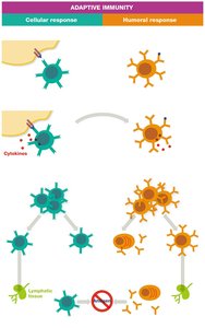
Adaptive immunity is a highly specific defense mechanism that involves the recognition and elimination of foreign antigens by specialized lymphocytes. The two main branches are the cellular response (mediated by T cells) and the humoral response (mediated by B cells and antibodies).
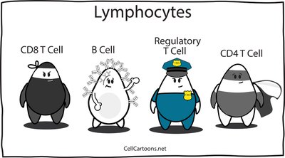
T and B Lymphocytes
Types and Development
T cells (CD8+ cytotoxic and CD4+ helper): Produced in the bone marrow, mature in the thymus.
B cells: Produced and mature in the bone marrow.
Both coordinate adaptive immune responses against foreign antigens.
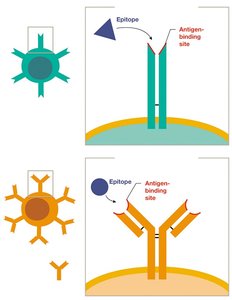
Antigen Receptors
T cell receptors (TCRs) and B cell receptors (BCRs) are specialized proteins that recognize specific epitopes on antigens.
Each lymphocyte expresses thousands of identical receptors, but each cell recognizes only one epitope type.
The diversity of T and B cells allows the immune system to recognize virtually any antigen.
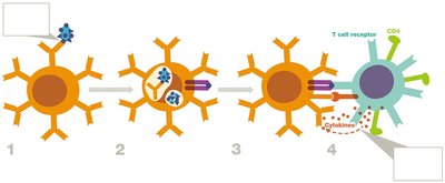
Stages of the Adaptive Immune Response
The adaptive immune response proceeds through four main stages:
Antigen Presentation
Lymphocyte Activation
Lymphocyte Proliferation and Differentiation
Antigen Elimination and Memory
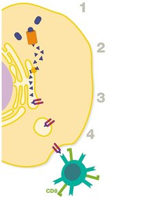
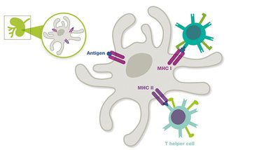
Stage 1: Antigen Presentation
Antigen-presenting cells (APCs) such as dendritic cells, macrophages, and B cells process and present antigens to T cells.
APCs use Major Histocompatibility Complex (MHC) molecules to display antigen fragments:
MHC I: Present on all nucleated cells; presents intracellular antigens to CD8+ cytotoxic T cells.
MHC II: Present only on APCs; presents extracellular antigens to CD4+ helper T cells.
Type | MHC I | MHC II |
|---|---|---|
Location | All nucleated cells (except RBCs) | APCs only |
Interacts with | CD8+ T cells | CD4+ T cells |
Antigens Presented | Intracellular | Extracellular |
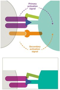
Stage 2: Lymphocyte Activation
T cells require two signals for activation:
Primary: TCR binds to MHC-antigen complex.
Secondary: Co-stimulatory proteins (e.g., B7 on APC binds CD28 on T cell).
B cells can be activated by direct antigen binding (T-independent) or require T helper cell assistance (T-dependent).
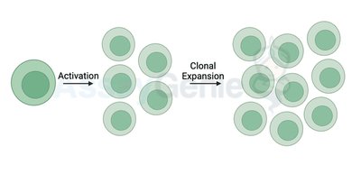
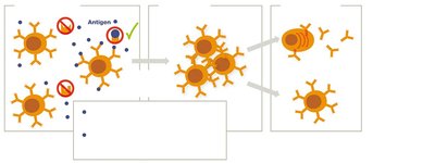
Stage 3: Lymphocyte Proliferation and Differentiation
Activated lymphocytes undergo clonal expansion (proliferation) and differentiate into effector and memory cells.
Effector cells: Actively participate in eliminating the antigen.
Memory cells: Remain in lymphatic tissues for rapid response upon re-exposure.
Stage 4: Antigen Elimination and Memory
Effector cells eliminate the antigen through cellular and humoral mechanisms.
After the threat is cleared, most effector cells die, but memory cells persist for years, providing immunological memory.
T Helper Cell Subsets and Functions
TH1 cells: Promote cellular responses by activating cytotoxic T cells, macrophages, and NK cells.
TH2 cells: Stimulate B cells to produce antibodies (humoral response).
T regulatory (Treg) cells: Suppress immune responses to maintain tolerance and prevent autoimmunity.
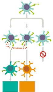
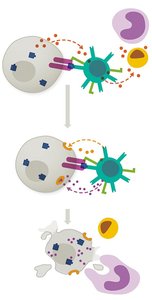
Cytotoxic T Cells (CD8+)
Directly destroy infected, damaged, foreign, or cancerous cells.
Mechanism: Release perforins (form pores) and granzymes (induce apoptosis) upon recognition of MHC I-antigen complex.
Humoral Immunity: B Cells and Antibodies
B Cell Activation
T-independent activation: Certain antigens (e.g., polysaccharides) can activate B cells directly.
T-dependent activation: Protein antigens require help from T helper cells (especially TH2) for full B cell activation.
Activation involves antigen binding to BCR and co-stimulatory interactions (e.g., CD40 on B cell with CD40L on T cell).
B Cell Proliferation and Differentiation
Activated B cells proliferate and differentiate into:
Plasma cells: Secrete antibodies specific to the antigen.
Memory B cells: Provide long-term immunity.
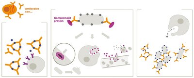
Antibody Functions
Antibodies (immunoglobulins) help eliminate antigens by:
Neutralizing toxins and pathogens
Activating complement cascade (leading to cytolysis, inflammation, opsonization)
Enhancing phagocytosis (precipitation, agglutination, opsonization)
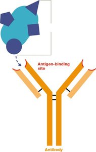
Antibody Structure and Isotypes
Basic structure: Two heavy chains, two light chains, antigen-binding sites at the tips.
Fc region determines the antibody's class and function.
Isotype switching allows B cells to produce different classes of antibodies with the same antigen specificity.
Isotype | Structure | Proportion | Neutralization | Complement Activation | Opsonization | Agglutination/Precipitation | Half-life | Notes |
|---|---|---|---|---|---|---|---|---|
IgG | Monomer | Most abundant | Strong | Strong | Strong | Strong | 21 days | Crosses placenta |
IgA | Monomer or dimer | Second most abundant | Strong | Some | Some | Negligible | 6 days | Main antibody in mucous |
IgM | Monomer or pentamer | Third most abundant | Strong | Strong | Negligible | Strong | 10 days | First made in infection |
IgE | Monomer | Rare | Negligible | Negligible | Negligible | Negligible | 2 days | Allergy/parasite responses |
IgD | Monomer | Rare | Negligible | Negligible | Negligible | Negligible | 2 days | B cell receptor; poorly understood |
Immunological Memory
Memory cells (both T and B) persist in lymphoid tissues after the initial response.
Upon re-exposure to the same antigen, memory cells mount a faster and stronger response (secondary response).
Example: The secondary antibody response is more rapid and robust than the primary response, providing the basis for long-lasting immunity after infection or vaccination.
*Additional info: The notes above integrate and expand upon the provided lecture slides, including definitions, mechanisms, and clinical relevance for adaptive immunity, T and B cell biology, and antibody function, as expected in a college-level microbiology course.*
