Back
Backchapter 18 Applications of Immunology: Vaccines and Serological Testing
Study Guide - Smart Notes
 Tailored notes based on your materials, expanded with key definitions, examples, and context.
Applications of Immunology
History of Vaccines and Herd Immunity
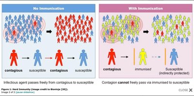
Vaccination is a foundational practice in immunology, involving the introduction of antigens to stimulate an immune response and confer protection against infectious diseases. The concept of herd immunity describes how immunizing a significant portion of a population can protect individuals who are not immune by reducing the overall spread of a pathogen.
Vaccination: The process of introducing a harmless form of a pathogen or its components to stimulate immunity without causing disease.
Jenner's Experiment: Edward Jenner used cowpox virus to immunize against smallpox, demonstrating cross-reactivity of antibodies.
Herd Immunity: Achieved when a large percentage of the population is immune, thereby limiting the spread of disease and protecting unimmunized individuals.
Reservoir Elimination: Herd immunity can eliminate the reservoir of infection, reducing outbreaks.
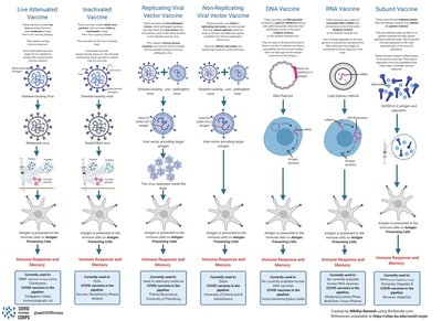
Types of Vaccines
Vaccines are classified based on their composition and the method by which they stimulate the immune system. Each type has specific advantages and applications.
Attenuated Whole-Agent Vaccines: Contain live, weakened pathogens (e.g., MMR vaccine).
Inactivated Whole-Agent Vaccines: Contain killed pathogens (e.g., Salk polio vaccine).
Toxoids: Inactivated bacterial toxins used to induce immunity (e.g., tetanus vaccine).
Subunit Vaccines: Contain only parts of the pathogen, such as proteins (e.g., acellular pertussis, recombinant hepatitis B).
Nucleic Acid (DNA/RNA) Vaccines: Contain genetic instructions for antigen production (e.g., West Nile vaccine for horses, COVID-19 mRNA vaccines).
Conjugate Vaccines: Polysaccharides conjugated with proteins to enhance immunogenicity (e.g., Hib, pneumococcal vaccines).
Principal Vaccines Used in the United States
Several vaccines are routinely used in the U.S. to prevent both bacterial and viral diseases. These vaccines have significantly reduced the incidence of many infectious diseases.
Bacterial Diseases: DTaP (diphtheria, tetanus, pertussis), meningococcal, Hib, pneumococcal vaccines.
Viral Diseases: Smallpox (no longer used), polio (Salk and Sabin), rabies, hepatitis A and B, influenza, MMR (measles, mumps, rubella), chickenpox.
Serological Tests
Overview of Serological Testing
Serological tests detect the presence of antibodies or antigens in a patient’s sample, providing valuable information for diagnosis, immunity status, and epidemiological studies.
Agglutination: Detects particulate antigens by clumping (used for antibody titers).
Precipitation: Detects soluble antigens by forming antigen-antibody complexes.
Fluorescent-Antibody Technique: Uses antibodies linked to fluorescent dyes for pathogen detection.
Complement Fixation: Detects antibody presence by measuring complement activation.
Hemagglutination: Agglutination of red blood cells, often used in blood typing and viral assays.
ELISA (Enzyme-Linked Immunosorbent Assay): Uses enzyme-linked antibodies to detect antigens or antibodies via color change.
Monoclonal Antibodies (Mabs)
Monoclonal antibodies are identical antibodies produced by a single clone of B cells, engineered for specificity to a single epitope. They are produced using hybridoma technology and have numerous diagnostic and therapeutic applications.
Production: Involves fusing an antibody-producing B cell with a myeloma (cancerous) cell to create a hybridoma capable of continuous growth and antibody production.
Applications: Used in treatments for transplant rejection (Muromonab-CD3), autoimmune diseases (Infliximab, Rituximab), cancer (Trastuzumab/Herceptin), and diagnostics.

Steps in Monoclonal Antibody Production
A mouse is immunized with a specific antigen to stimulate antibody production.
The spleen is removed and homogenized to obtain B cells producing the desired antibody.
Spleen cells are fused with myeloma cells to form hybridomas.
The mixture is placed in selective medium; only hybridomas survive and proliferate.
Hybridomas are screened for production of the desired antibody.
Selected hybridomas are cultured to produce large quantities of monoclonal antibodies.
Precipitation and Agglutination Reactions
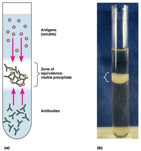
Precipitation Reactions
Precipitation reactions occur when soluble antigens react with antibodies to form visible complexes. The precipitin ring test is a classic example, used to quantify antigen-antibody interactions.
Zone of Equivalence: The optimal ratio of antigen to antibody where maximal precipitation occurs.
Applications: Used in diagnostic immunology to detect and quantify specific antigens or antibodies.
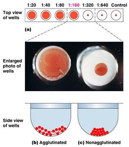
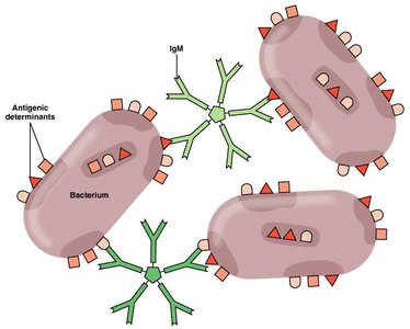
Agglutination Reactions
Agglutination involves the clumping of particles, such as cells or latex beads, when antibodies bind to antigens on their surfaces. This reaction is used to measure antibody titers and for blood typing.
Direct Agglutination: Antigen is naturally present on the cell surface (e.g., blood cells, bacteria).
Indirect (Passive) Agglutination: Antigen is attached to inert particles like latex beads.
Antibody Titer: The highest dilution of serum that still produces agglutination, indicating antibody concentration.
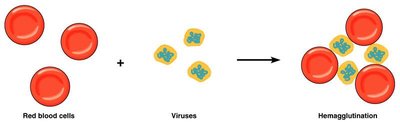
Hemagglutination and Neutralization Reactions
Hemagglutination is the clumping of red blood cells, often used to detect viruses that agglutinate RBCs. Neutralization reactions use antibodies to block the harmful effects of toxins or viruses.
Hemagglutination: Used in blood typing and viral assays.
Neutralization: Antibodies neutralize toxins or viruses, preventing them from damaging cells.
Fluorescent Antibody Techniques
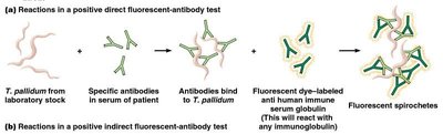
Direct and Indirect Fluorescent Antibody Tests
Fluorescent antibody techniques use antibodies labeled with fluorescent dyes to detect specific antigens or antibodies. These tests are highly sensitive and specific, commonly used in research and diagnostics.
Direct Test: Labeled antibody binds directly to the antigen on the pathogen.
Indirect Test: Patient serum is added to a fixed antigen; a secondary fluorescent antibody detects bound patient antibodies.
Enzyme-Linked Immunosorbent Assay (ELISA)
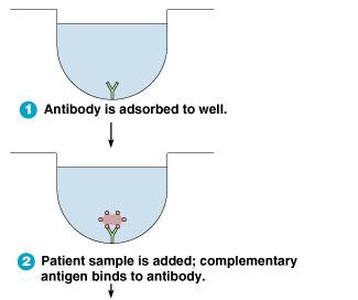
Direct ELISA
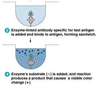
Direct ELISA detects the presence of antigens in a sample using enzyme-linked antibodies. A color change indicates a positive reaction, making this test useful for pathogen detection.
Antibody is adsorbed to the well.
Patient sample is added; antigen binds to the antibody.
Enzyme-linked antibody specific for the antigen is added, forming a sandwich.
Enzyme substrate is added; a color change indicates a positive result.
Indirect ELISA
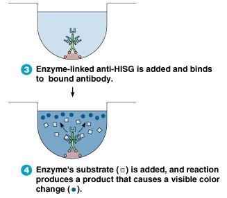
Indirect ELISA detects antibodies in a patient’s serum. The test uses an antigen-coated well, patient serum, and an enzyme-linked secondary antibody. A color change indicates the presence of specific antibodies.
Antigen is adsorbed to the well.
Patient serum is added; specific antibodies bind to the antigen.
Enzyme-linked anti-human immunoglobulin is added, binding to the patient antibody.
Enzyme substrate is added; a color change indicates a positive result.
Application: Home Pregnancy Test
Home pregnancy tests are a practical application of the sandwich ELISA principle. They detect human chorionic gonadotropin (hCG) in urine using monoclonal antibodies, producing a visible color change if hCG is present.
Capture Antibody: Immobilized on the test strip to bind hCG.
Free Antibody: Labeled with a color indicator, binds to hCG and forms a sandwich complex.
Result: Color change in the test window indicates pregnancy.