 Back
Backlecture 11
Study Guide - Smart Notes
 Tailored notes based on your materials, expanded with key definitions, examples, and context.
Bacterial Cell Properties
Motility
Bacterial motility refers to the ability of bacteria to move independently, often using specialized structures such as flagella. This movement is crucial for survival, colonization, and pathogenesis.
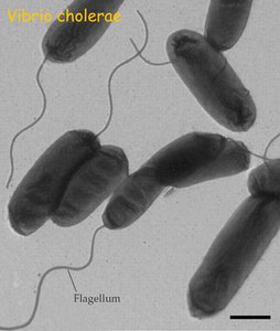
Flagellum: A helical filament composed of polymerized chains of the protein flagellin. Flagella are highly conserved among bacteria, making them targets for immune recognition (e.g., TLR-5 in vertebrates).
Types of Motility: Includes flagellar, twitching, and gliding motility. Some bacteria, like Listeria, use actin-based motility within host cells.
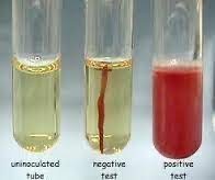
Motility Tests: Motility can be visualized and measured using soft agar media or microscopy.
Regulation: Pathogenic bacteria often regulate flagellin expression to evade immune detection inside the host.
Example: Vibrio cholerae uses a single polar flagellum for movement in both environmental water and host mucus. Non-motile mutants are less virulent.
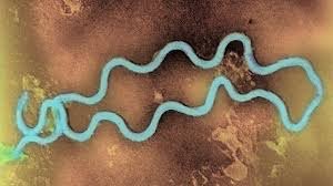
Spirochetes: Such as Treponema pallidum, possess a flagellum located in the periplasm, helping them evade immune detection.
E. coli: Typically have 4-10 flagella per cell. Counter-clockwise rotation aligns flagella for straight swimming, while clockwise rotation causes tumbling.
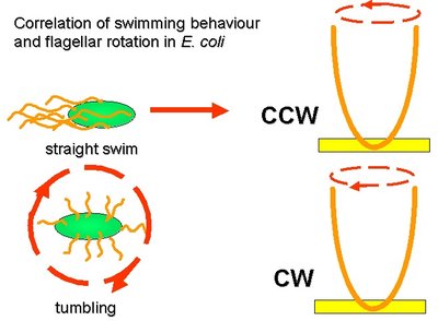
Random Walk Model: E. coli alternates between runs and tumbles, adjusting movement based on chemical gradients.

Motility Visualization: Motility can be tested in soft agar tubes; non-motile bacteria remain at the inoculation site, while motile bacteria spread.
Chemotaxis and Other Taxes
Chemotaxis is the directed movement of bacteria in response to chemical gradients. Other forms of taxes include aerotaxis (oxygen), phototaxis (light), magnetotaxis (magnetic fields), osmotaxis (salinity), and hydrotaxis (water).
Mechanism: Bacteria sense attractants or repellents and adjust their movement accordingly.
Role in Virulence: Chemotaxis is essential for pathogens to locate and colonize host tissues.
Measurement: Chemotaxis can be measured using capillary tubes containing attractants or repellents, or observed microscopically.
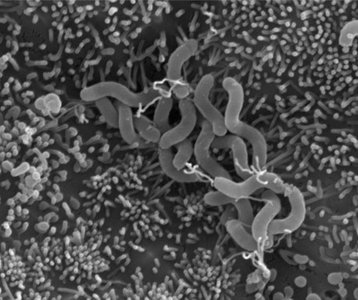
Example: Helicobacter pylori uses chemotaxis to penetrate deep into gastric glands.
Cell-to-Cell Spread: Listeria monocytogenes
Actin-Based Motility
Listeria monocytogenes is a Gram-positive, food-borne pathogen capable of intracellular movement using host cell actin. This mechanism allows rapid cell-to-cell spread, avoiding exposure to the humoral immune system.
Pathogenesis: Listeria can cause severe disease, especially in immunocompromised individuals, and can cross the placenta.
Growth at Low Temperatures: Listeria is a psychrophile, able to grow at refrigeration temperatures.
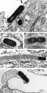
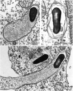
Actin Comet Tails: Listeria hijacks host actin to propel itself through the cytosol.


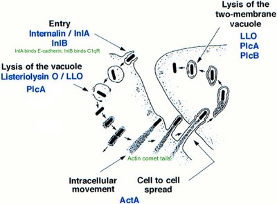
Immune Evasion: Intracellular spread allows Listeria to avoid antibody-mediated immunity.
Adhesion and Colonization
Bacterial Adhesion Mechanisms
Adhesion is the process by which bacteria attach to surfaces, cells, or tissues, a critical step for colonization and pathogenesis.
Adhesins: Cell-surface proteins that facilitate attachment to host cells or surfaces.
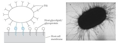
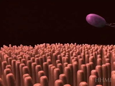
Pili/Fimbriae: Long, rod-like structures with adhesins at their tips, mediating initial attachment.
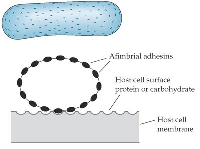
Afimbrial Adhesins: Surface proteins that mediate tight binding to host cell proteins, often involved in intimate attachment.
Fibrillar Adhesins: Short, thin rods binding to extracellular matrix proteins (fibronectin, collagen, etc.).
Attachment Process: Usually a two-step process: loose attachment (pili/fimbriae/fibrillar adhesins) followed by tight attachment (afimbrial adhesins).
Tissue Tropism: Specific adhesion leads to preference for certain tissues or surfaces.
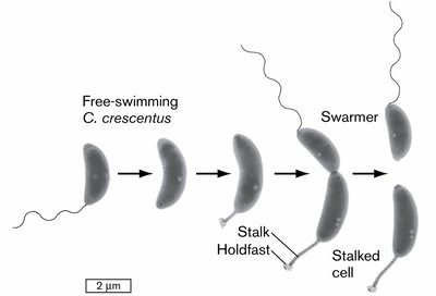
Example: Caulobacter crescentus undergoes asymmetric cell division, producing stationary and mobile daughter cells. The holdfast binds to surfaces, while the flagellum allows swimming.
Colonization: Adherence/Attachment
Colonization begins with adherence to mucosal or environmental surfaces, often in a two-step process. Bacteria capable of adhering to mucosal cells have a survival advantage.
Gram-negative Adhesins: Pili/fimbriae are helical arrays of pilin protein, often with a tip protein for host receptor binding.
Afimbrial Adhesins: Mediate tight binding, often to host surface proteins rather than sugars.
Gram-positive Adhesins: Fibrillar adhesins bind to connective tissue and extracellular matrix proteins.
Pathogen Strategies: Some pathogens inject additional adhesin proteins into host cells to enhance binding and modulate host responses.
Summary Table: Bacterial Adhesion Structures
Structure | Type | Function | Example |
|---|---|---|---|
Pili/Fimbriae | Gram-negative | Loose attachment, initial binding | E. coli |
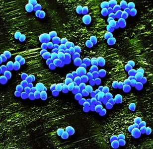
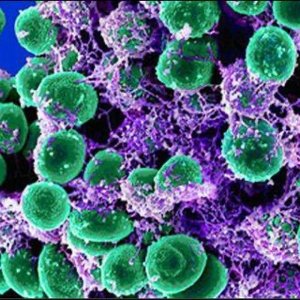
Afimbrial Adhesins | Both | Tight attachment, intimate binding | Staphylococcus |
Fibrillar Adhesins | Gram-positive | Binding to extracellular matrix | Streptococcus |
Holdfast | Environmental | Surface binding, asymmetric division | Caulobacter crescentus |
Example: Pathogenic E. coli colonization involves a two-step attachment: loose (pili/fimbriae) and tight (afimbrial adhesins).
Additional info: Some pathogens further modify adherence by injecting effector proteins into host cells, enhancing colonization and modulating host cell responses.
