 Back
Backlec 05:Bacterial Cell Structure and Function: Cell Envelope, Wall, Capsule, and DNA Replication
Study Guide - Smart Notes
 Tailored notes based on your materials, expanded with key definitions, examples, and context.
Bacterial Cell Structure and Function
Overview of Bacterial Cells
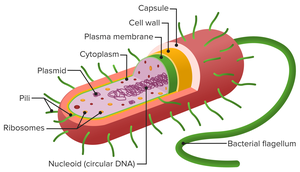
Bacterial cells are microscopic, single-celled prokaryotes characterized by the absence of a nucleus and membrane-bound organelles. Their structural organization is fundamental to their survival, reproduction, and pathogenicity.
Cell Envelope: Multilayered structure providing shape and protection.
Nucleoid: Region containing circular DNA.
Reproduction: Primarily via binary fission.
Metabolic Diversity: Bacteria exhibit a wide range of metabolic capabilities.
Bacterial Cell Envelope
Key Structural Components and Functions
The bacterial cell envelope is essential for maintaining cell shape, protecting against environmental stressors, and mediating interactions with the environment.
Capsule: Outermost layer, often composed of polysaccharides.
Cell Wall: Provides structural strength.
Plasma Membrane: Regulates transport and metabolic processes.
The Capsule: Structure and Function
Composition and Core Functions
The capsule is a protective layer that enhances bacterial survival and virulence.
Protection: Acts as a barrier against toxic compounds, prevents desiccation, and shields from host immune defenses.
Adhesion: Promotes attachment to surfaces and facilitates biofilm formation.
Immune Evasion Mechanisms
Encapsulated bacteria employ several strategies to evade host immunity:
Anti-Phagocytic Shield: The capsule's slippery surface impedes phagocytosis by immune cells.
Molecular Mimicry: Capsules may contain molecules found in human tissues, causing immune misidentification.

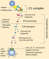
Blocking Complement Activation
The capsule masks binding sites required for complement activation, preventing formation of the membrane attack complex.
Complement System: Blood proteins that bind bacterial surfaces and form membrane-destroying pores.
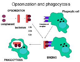
Hindering Opsonization
Capsules conceal surface antigens, reducing antibody binding and subsequent immune clearance.
Opsonization: Antibodies tag bacteria for destruction; capsules interfere with this process.
Clinical Importance of Capsules
Encapsulated bacteria, such as Streptococcus pneumoniae and Neisseria meningitidis, are often more virulent and cause severe invasive diseases.
The Cell Wall: Structural Strength and Composition
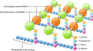
Peptidoglycan Structure
The bacterial cell wall is a mesh-like structure composed of peptidoglycan, providing mechanical strength and shape.
Sugar Backbone: Alternating units of N-acetylglucosamine (NAG) and N-acetylmuramic acid (NAM).
Peptide Cross-Links: Short peptide chains form covalent links between glycan strands.
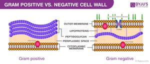
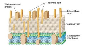
Gram-Positive Cell Wall
Gram-positive bacteria have a thick, multilayered peptidoglycan wall reinforced by teichoic acids.
Thickness: 30–100 layers of peptidoglycan.
Teichoic Acids: Impart negative charge, bind cations, and reinforce wall stability.

Gram-Negative Cell Wall
Gram-negative bacteria possess a thin peptidoglycan layer within the periplasmic space and an outer membrane containing lipopolysaccharides (LPS).
Outer Membrane: Acts as a defensive barrier; contains LPS, which functions as an endotoxin.
Periplasm: Gel-like compartment rich in enzymes and transport proteins.
Porins: Protein channels allowing selective entry of small molecules.

Osmotic Lysis and Cell Wall Function
Protection Against Osmotic Pressure
Bacteria inhabit hypotonic environments, where water influx could cause cell rupture. The peptidoglycan mesh prevents osmotic lysis by counteracting internal turgor pressure.
Mechanical Strength: Withstands pressures up to ~20 atmospheres.
Penicillin and Cell Wall Synthesis
Mechanism of Action
Penicillin targets penicillin-binding proteins (PBPs), inhibiting transpeptidation and preventing proper cell wall construction.
Molecular Mimicry: Penicillin mimics the D-Ala–D-Ala motif, irreversibly inactivating PBPs.
Result: Weak, unstable peptidoglycan layers lead to osmotic lysis.
Gram Staining Technique
Principle and Steps
Gram staining differentiates bacteria based on cell wall structure and is a primary diagnostic tool.
Primary Stain: Crystal violet stains all cells purple.
Mordant: Gram’s iodine forms a CV–I complex.
Decolorization: Alcohol removes dye from Gram-negative cells.
Counterstain: Safranin stains Gram-negative cells pink/red.
Bacterial DNA Replication
Overlapping Replication Cycles
Bacteria initiate new rounds of DNA replication before previous cycles finish, enabling rapid growth.
Origin of Replication (oriC): Site where replication begins.
Nested Replication: Multiple replication forks operate simultaneously.
Biological Trade-offs
Increased Mutation Pressure: Rapid replication elevates mutation rates.
High Metabolic Demand: Requires continuous supply of nucleotides and ATP.
Ciprofloxacin: Disrupting DNA Replication
Mechanism of Action
Ciprofloxacin targets DNA gyrase and topoisomerase IV, enzymes essential for relieving DNA supercoiling and separating chromosomes.
Enzyme–DNA Trapping: Stabilizes the DNA–enzyme cleavage complex, preventing re-ligation.
Replication Fork Collision: Leads to double-stranded DNA breaks and catastrophic chromosomal damage.
SOS Response: Error-prone repair mechanisms are activated, often resulting in cell death.
Summary Table: Gram-Positive vs. Gram-Negative Cell Wall
Feature | Gram-Positive | Gram-Negative |
|---|---|---|
Peptidoglycan Thickness | Thick (30–100 layers) | Thin (1–3 layers) |
Teichoic Acids | Present | Absent |
Outer Membrane | Absent | Present (contains LPS) |
Periplasmic Space | Minimal | Prominent |
Drug Resistance | Lower | Higher |
