 Back
BackBacterial Cell Structure and Function: Cell Walls, Membranes, and Cytoplasm
Study Guide - Smart Notes
 Tailored notes based on your materials, expanded with key definitions, examples, and context.
Cell Structure and Function
Overview
This section explores the fundamental structures and functions of bacterial cells, focusing on the cell wall, cytoplasmic membrane, and cytoplasm. Understanding these components is essential for classifying bacteria, understanding their physiology, and targeting them with antibiotics.
Cell Wall
Functions of the Cell Wall
Protection: The cell wall protects the cytoplasmic membrane and provides structural support, preventing osmotic lysis (bursting due to water influx).
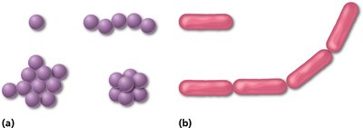
Shape: It gives bacteria their characteristic shapes (e.g., cocci, bacilli).
Attachment and Resistance: Some cell walls assist in attachment to other cells or surfaces and help resist antimicrobial drugs.
Antibiotic Target: Many antibiotics target the bacterial cell wall, making it a critical structure in medical microbiology.
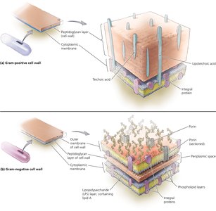
Composition and Types
Peptidoglycan: The main component of bacterial cell walls, consisting of sugar chains cross-linked by peptides.
Two Major Types:
Gram-positive: Thick peptidoglycan layer, teichoic acids, no outer membrane.
Gram-negative: Thin peptidoglycan layer, outer membrane with lipopolysaccharides (including Lipid A).
Pathogenicity: Cell wall components contribute to a bacterium's ability to cause disease.
Diagnostic Importance: Differences in cell wall structure are the basis for the Gram stain, a key diagnostic tool.
Gram Staining and Acid-Fast Staining
Gram Stain: Differentiates bacteria based on cell wall structure.
Gram-positive: Stain purple (thick peptidoglycan retains crystal violet dye).
Gram-negative: Stain pink (thin peptidoglycan does not retain dye after alcohol wash).
Acid-Fast Stain: Identifies bacteria with large amounts of mycolic acid in their cell wall (e.g., Mycobacteria).
Feature | Gram-Positive | Gram-Negative |
|---|---|---|
Gram Stain | Purple | Pink |
Peptidoglycan Layer | Thick | Thin |
Teichoic Acids | Present | Absent |
Outer Membrane | Absent | Present |
Lipopolysaccharides | Absent | Present (Lipid A) |
Flagella Basal Body | Two rings | Four rings |
Cytoplasmic Membrane
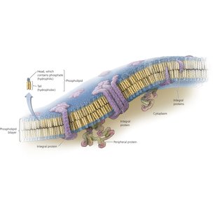
Structure
Phospholipid Bilayer: Composed of hydrophilic phosphate heads facing outward and hydrophobic fatty acid tails inward.
Proteins: Integral and peripheral proteins are embedded in or associated with the membrane, serving various functions.
Function
Selective Permeability: Controls the passage of substances into and out of the cell.
Barrier: Naturally impermeable to most substances; proteins facilitate transport.
Gradients: Maintains concentration and electrical gradients essential for cellular processes.
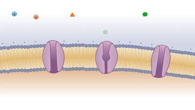
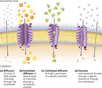
Movement Across the Membrane
Passive Processes: Do not require energy; substances move down their electrochemical gradient.
Diffusion: Movement of small or lipid-soluble molecules (e.g., O2, CO2).
Facilitated Diffusion: Movement via channels or carrier proteins (e.g., glucose, urea).
Osmosis: Movement of water across a selectively permeable membrane.
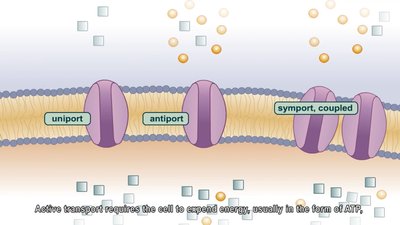
Active Processes: Require energy (ATP); substances move against their gradient.
Active Transport: ATP-dependent carrier proteins transport ions (e.g., Na+, K+).
Group Translocation: Substance is chemically modified during transport (e.g., glucose phosphorylation).
Transport Type | Description | Examples of Substances |
|---|---|---|
Diffusion | Molecules move down gradient through bilayer | O2, CO2, lipid-soluble chemicals |
Facilitated Diffusion | Molecules move down gradient via proteins | Glucose, urea, some vitamins |
Osmosis | Water moves down gradient | Water |
Active Transport | ATP-dependent carrier proteins move substances against gradient | Na+, K+, Ca2+, H+, Cl- |
Group Translocation | Substance chemically altered during transport | Glucose, mannose, fructose |
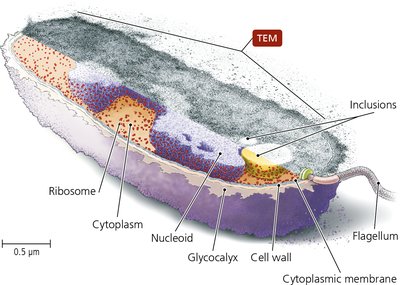
Cytoplasm
Components
Cytosol: The liquid portion, mainly water, containing dissolved ions, nutrients, and proteins.
Nucleoid: Region containing the bacterial chromosome (DNA).
Inclusions: Reserve deposits of chemicals (e.g., glycogen, polyphosphate granules).
Endospores: Dormant, highly resistant structures formed by some bacteria for survival in harsh conditions.
Ribosomes: Sites of protein synthesis; bacterial ribosomes are 70S in size.
Cytoskeleton: Protein filaments that help maintain cell shape and assist in cell division.
Key Terms and Concepts
Carrier Proteins: Used in facilitated diffusion, a form of passive transport.
Gram Staining: Targets differences in cell wall structure to classify bacteria as Gram-positive or Gram-negative.
Acid-Fast Staining: Identifies bacteria with mycolic acid-rich cell walls, such as Mycobacteria.
Summary Table: Bacterial Cell Structure and Function
Structure | Main Components | Function |
|---|---|---|
Cell Wall | Peptidoglycan, teichoic acids (G+), LPS (G-) | Shape, protection, antibiotic target |
Cytoplasmic Membrane | Phospholipid bilayer, proteins | Selective barrier, transport, gradients |
Cytoplasm | Cytosol, nucleoid, inclusions, ribosomes, cytoskeleton | Metabolism, genetic material, storage, protein synthesis |
Example Application: The Gram stain is routinely used in clinical microbiology to rapidly identify bacterial pathogens and guide antibiotic therapy.
