 Back
Bacterial Staining Techniques and Cell Wall Structure
Study Guide - Smart Notes
 Tailored notes based on your materials, expanded with key definitions, examples, and context.
Bacterial Staining
Introduction to Staining
Staining is a fundamental technique in microbiology used to enhance the visibility and contrast of microorganisms under the microscope. Most bacteria are naturally transparent and colorless, making them difficult to observe without staining. Stains are charged, colored compounds that interact with bacterial cells to provide contrast.
Basic stains (cationic): Positively charged, attracted to negatively charged bacterial cells. Examples: crystal violet, methylene blue, safranin, carbol fuchsin.
Acidic stains (anionic): Negatively charged, repelled by bacterial cells. Example: picric acid.
Purpose: Improves contrast, enhances visualization of cell morphology and arrangement.
Preparation of Bacterial Smear
Smear Preparation Process
A bacterial smear is a thin film of a solution of microbes spread onto a glass slide. Proper smear preparation is essential for successful staining and observation.
Air drying: Preserves cell integrity.
Heat fixing: Adheres cells to the slide and kills them, preventing wash-off during staining.
Quality: A good smear is thin and translucent; thick smears hinder light passage and obscure cell morphology.
Simple Staining
Principle and Procedure
Simple staining involves the application of a single basic stain to a bacterial smear. This technique allows for the visualization of cell shape and arrangement.
Procedure: Apply stain, wait one minute, rinse, dry, and observe.
Mordant: May be used to enhance stain retention or enlarge cells.
Visualization: Reveals cell shapes (cocci, bacilli, spirilla) and arrangements (chains, clusters, pairs, tetrads).
Negative Staining
Principle and Advantages
Negative staining is used to observe cellular morphology without distorting the cells. The stain (e.g., nigrosin) colors the background, leaving cells unstained and clearly visible.
High contrast: Cells appear bright against a dark background.
No distortion: Cell morphology is preserved.
Differential Staining
Gram Stain
The Gram stain is a differential staining technique that separates bacteria into two groups based on cell wall structure: Gram-positive and Gram-negative. It uses two contrasting stains and a series of steps to distinguish these groups.
Gram-positive: Thick peptidoglycan layer (~90% of cell wall), many pores.
Gram-negative: Thin peptidoglycan layer (~10% of cell wall), outer membrane with lipopolysaccharide (LPS), contains Lipid A (endotoxin).
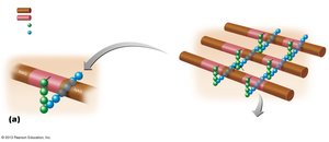
Structure of Peptidoglycan
Peptidoglycan is a complex polymer forming the bacterial cell wall, especially prominent in Gram-positive bacteria. It consists of repeating units of N-acetylglucosamine (NAG) and N-acetylmuramic acid (NAM), cross-linked by peptide bridges.
Carbohydrate backbone: Alternating NAG and NAM units.
Peptide cross-bridges: Provide structural strength.
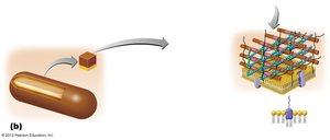
Gram-Positive Cell Wall
Gram-positive bacteria have a thick peptidoglycan layer and may contain teichoic acids, which contribute to cell wall rigidity and function.
Teichoic acids: Wall teichoic acid and lipoteichoic acid.
Absence of outer membrane: Unlike Gram-negative bacteria.
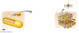
Gram-Negative Cell Wall
Gram-negative bacteria possess a thin peptidoglycan layer and an outer membrane containing lipopolysaccharide (LPS), which includes Lipid A, a potent endotoxin.
Outer membrane: Contains LPS, phospholipids, and proteins.
Periplasmic space: Located between the plasma membrane and outer membrane.
Comparative Characteristics of Gram-Positive and Gram-Negative Bacteria
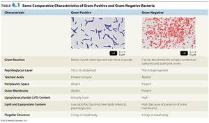
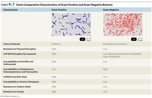
The following tables summarize the structural and functional differences between Gram-positive and Gram-negative bacteria.
Characteristic | Gram-Positive | Gram-Negative |
|---|---|---|
Gram Reaction | Retains crystal violet stain (purple) | Decolorized and counterstained (red/pink) |
Peptidoglycan Layer | Thick, multilayered | Thin, single layered |
Teichoic Acid | Present | Absent |
Periplasmic Space | Absent | Present |
Outer Membrane | Absent | Present |
Lipopolysaccharide (LPS) Content | None | High |
Flagellar Structure | 2 rings in basal body | 4 rings in basal body |
Characteristic | Gram-Positive | Gram-Negative |
|---|---|---|
Toxins Produced | Exotoxins | Endotoxins and exotoxins |
Resistance to Physical Disruption | High | Low |
Cell Wall Disrupting Agents | High | Low |
Susceptibility to Penicillin | High | Low |
Susceptibility to Basic Dyes | High | Low |
Resistance to Sodium Azide | High | Low |
Resistance to Drying | High | Low |
Gram Staining Procedure
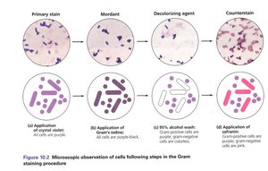
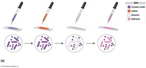
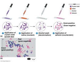
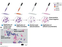
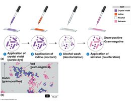
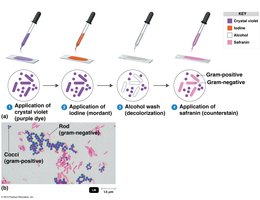
The Gram staining process involves four main steps, each with a specific purpose:
Primary stain (crystal violet): Stains all cells purple.
Mordant (iodine): Forms a complex with crystal violet, enhancing retention.
Decolorization (alcohol): Removes stain from Gram-negative cells.
Counterstain (safranin): Stains Gram-negative cells pink/red.

Microscopic Appearance After Gram Staining
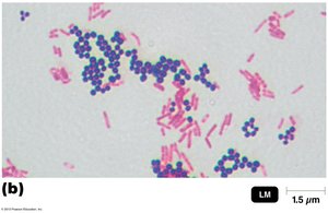
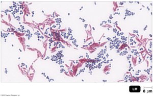
After Gram staining, Gram-positive bacteria appear purple, while Gram-negative bacteria appear pink/red. The morphology and arrangement of cells can be observed.
Differential Staining: Acid-Fast Stain
Principle and Procedure
The acid-fast stain is used to identify bacteria of the genus Mycobacterium, which have a waxy cell wall containing mycolic acid. These cells resist decolorization by acid alcohol and retain the primary stain (carbol fuchsin).
Primary stain: Carbol fuchsin (red).
Heat: Softens waxy cell wall for stain penetration.
Decolorization: Acid alcohol removes stain from non-acid-fast cells.
Counterstain: Methylene blue stains non-acid-fast cells blue.
Result: Acid-fast cells are red; non-acid-fast cells are blue.

Applications and Significance
Acid-fast staining is crucial for diagnosing diseases caused by Mycobacterium species, such as tuberculosis and leprosy. The waxy cell wall also confers resistance to phagocytosis and many antibiotics.
Diagnostic value: Identifies pathogenic acid-fast bacteria.
Clinical relevance: Resistance to antibiotics and immune defenses.
