 Back
Bacteriophages and Viral Specificity: Lab Study Guide
Study Guide - Smart Notes
 Tailored notes based on your materials, expanded with key definitions, examples, and context.
Viruses: General Characteristics
Living and Non-Living Characteristics
Viruses are unique infectious agents exhibiting both living and non-living properties. They are central to microbiology due to their role in disease and genetic exchange.
Living characteristics: Viruses reproduce rapidly, but only within living host cells, and can mutate.
Non-living characteristics: Viruses are acellular, lack cytoplasm and organelles, cannot carry out metabolism independently, and replicate using host cell machinery. They possess either DNA or RNA, but never both.
Size Comparison: Viruses vs. Bacteria
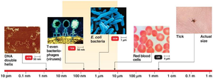
Viruses are much smaller than bacteria, typically ranging from 10–250 nm, making them submicroscopic and invisible to light microscopy.
Bacteria: Usually 1–10 µm in size.
Viruses: 10–250 nm, requiring electron microscopy for visualization.
Structure of Viruses
Basic Components
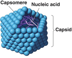
Viruses are structurally simpler than bacteria, consisting mainly of a genome and a protein coat.
Genome: Either DNA or RNA, single or double-stranded.
Capsid: Protein shell composed of subunits called capsomeres.
Naked vs. Enveloped Viruses
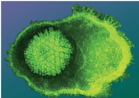
Naked viruses: Consist only of nucleic acid and capsid (nucleocapsid).
Enveloped viruses: Have an envelope derived from the host cell's plasma membrane surrounding the nucleocapsid.
Bacteriophages
Definition and Structure
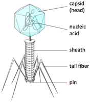
Bacteriophages are viruses that specifically infect bacteria. Some possess complex tail structures for attachment and genome injection.
Head (capsid): Contains nucleic acid.
Tail: Used for attachment and genome delivery.
Bacteriophage Replication: The Lytic Cycle
Stages of the Lytic Cycle
The lytic cycle is the primary mode of bacteriophage replication, resulting in destruction of the host cell.
Attachment: Bacteriophage tail fibers bind to specific receptor sites on the bacterial cell wall.
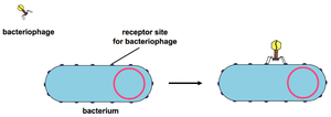
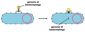
Penetration: Lysozyme creates a hole in the cell wall; the phage injects its genome into the bacterium.
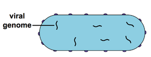
Biosynthesis: Host macromolecular synthesis is shut down; phage genome replicates and directs synthesis of viral components.
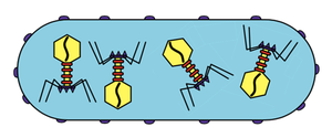
Maturation: Newly synthesized phage parts assemble into complete virions.
Release: Lysozyme breaks down peptidoglycan, causing osmotic lysis and release of new phages.
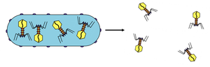
Reinfection: 50–200 new phages infect surrounding bacteria, continuing the cycle.
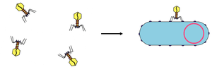
Viral Specificity and Phage Typing
Viral Specificity
Viral specificity refers to the precise interaction between a bacteriophage and its host bacterium, similar to enzyme-substrate or antibody-antigen specificity.
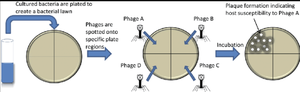
Phage typing: Using known phages to identify unknown bacteria based on susceptibility and lysis.
Phage Typing Procedure
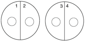
Divide agar plates into sectors for different bacterial samples.
Streak unknown bacteria in each sector.
Add a drop of phage suspension to each sector.
Incubate and observe for lysis (clear zones).
Plaque Formation and Counting
Plaques and Plaque Counting
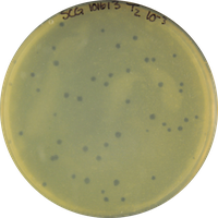
A plaque is a clear area on an agar plate where bacteria have been lysed by phages. Plaque counting is used to quantify phage concentration.
Plaque Forming Units (PFU): Each plaque represents one infectious phage particle.
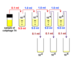
Plaque count procedure: Serial dilution of phage suspension, mixing with host bacteria, plating, and counting plaques.
Calculating PFUs
To determine PFUs per milliliter:
Count plaques on a plate (ideally 30–300).
Multiply by the dilution factor of the plate.
Formula: $\text{PFU/ml} = \text{Number of plaques} \times \text{Dilution factor}$
Summary Table: Virus and Bacteriophage Characteristics
Feature | Viruses | Bacteriophages |
|---|---|---|
Host Range | Animals, plants, bacteria | Bacteria only |
Genome | DNA or RNA | DNA (usually) |
Structure | Capsid, sometimes envelope | Capsid, tail fibers, sheath |
Replication | Requires host cell | Lytic or lysogenic cycle |
Identification | Serology, PCR, etc. | Phage typing |
Key Terms and Concepts
Capsid: Protein shell of a virus.
Capsomere: Subunit of the capsid.
Nucleocapsid: Combination of nucleic acid and capsid.
Envelope: Lipid membrane surrounding some viruses.
Bacteriophage: Virus that infects bacteria.
Lytic cycle: Viral replication resulting in host cell lysis.
Plaque: Clear zone on agar plate indicating bacterial lysis.
Phage typing: Method for identifying bacteria using phage specificity.
PFU: Plaque Forming Unit, a measure of phage concentration.
