 Back
BackBIO 201 Unit 1: Introduction to Microbiology – Foundations, Classification, Microscopy, and Bioterrorism
Study Guide - Smart Notes
 Tailored notes based on your materials, expanded with key definitions, examples, and context.
Overview of Microbiology
Background of Microbiology
Microbiology is the scientific study of microorganisms, or microbes, which are forms of life too small to be seen with the naked eye. This field encompasses a diverse group of organisms, including bacteria, viruses, fungi, protozoa, algae, and helminths. Microbiologists use microscopes to observe and study these organisms, which play essential roles in health, ecology, and industry.
Microorganisms (Microbes): Include bacteria, viruses, fungi (yeasts, molds, mushrooms), protozoa, algae, and helminths.
Microscopy: Essential for visualizing microbes due to their small size.
Importance and Roles of Microbes
Not all microbes are harmful; many are beneficial and essential for life on Earth. They are part of the normal human microbiota, contribute to nutrient cycling, and are used in various industries.
Normal Flora: The human body contains more bacterial cells than human cells, with microbes aiding in digestion and acting as a barrier to pathogens.
Decomposition: Microbes decompose organic matter, increasing soil fertility.
Industrial Uses: Microbes are used in sewage treatment, food production (e.g., cheese, yogurt), and pharmaceuticals (antibiotics, vaccines).
Biotechnology: Microbes are tools in genetic engineering, gene therapy, and bioremediation (cleaning up pollutants).
Photosynthesis and Ecology: Many microbes contribute to photosynthesis and the recycling of elements in ecosystems.
Subdivisions of Microbiology
Bacteriology: Study of bacteria
Virology: Study of viruses
Mycology: Study of fungi
Parasitology: Study of protozoa and helminths
Immunology: Study of immune responses
Chemotherapy: Study of chemicals used to treat diseases
History of Microbiology
Early Observations and the Microscope
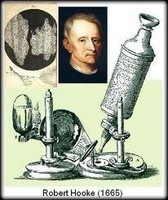
The development of the microscope was pivotal in the discovery of microorganisms. Robert Hooke and Anton van Leeuwenhoek made significant contributions to early microscopy and the observation of cells and bacteria.
Robert Hooke (1665): Developed the compound microscope and observed cells in cork.
Anton van Leeuwenhoek (1673): First to observe bacteria from teeth scrapings.
Origins of Life: Spontaneous Generation vs. Biogenesis
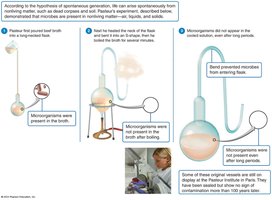
Early scientists debated whether life could arise spontaneously from non-living matter (spontaneous generation) or only from pre-existing life (biogenesis).
Spontaneous Generation (Abiogenesis): The belief that life arises from non-living matter.
Francesco Redi (1668): Demonstrated that maggots do not arise from meat unless flies lay eggs on it, supporting biogenesis.
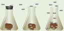
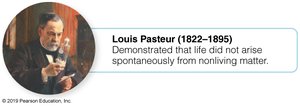
Louis Pasteur (1861): Disproved spontaneous generation with his swan-neck flask experiment, showing that microorganisms do not arise from non-living matter.
Major Contributions to Microbiology
Louis Pasteur: Developed aseptic techniques, rabies vaccine, discovered fermentation and pasteurization, and proposed the germ theory of disease.
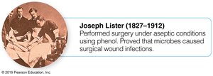
Joseph Lister (1867): Introduced disinfectants (phenol) in surgery, proving microbes cause surgical wound infections.
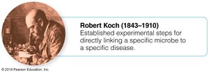
Robert Koch (1876): Established Koch’s postulates, linking specific microbes to specific diseases (e.g., Bacillus anthracis causes anthrax).
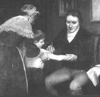
Edward Jenner (1798): Developed the first vaccination (smallpox), introducing the concept of immunity.
Elie Metchnikoff (1884): Discovered phagocytosis and cellular immunity.
Hans Christian Gram (1884): Developed the Gram stain for bacterial classification.
Richard Petri (1887): Invented the Petri dish for culturing microbes.
Dimitri Iwanowski (1892): Discovered viruses (Tobacco Mosaic Virus).
Paul Ehrlich (1910): Developed the first chemotherapeutic agent (“magic bullet”).
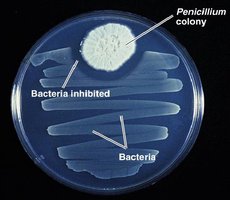
Alexander Fleming (1945): Discovered penicillin, the first antibiotic.
Selman Waksman (1952): Discovered streptomycin (used for TB) and coined the term “antibiotic.”
Rebecca Lancefield (1933): Classified Streptococcus species by serotypes.
Classification of Microorganisms
Taxonomy and the Five Kingdom System
Taxonomy is the science of classifying organisms. The five kingdom system, proposed by Robert Whittaker in 1969, categorized life into Monera, Protista, Fungi, Plantae, and Animalia.
Monera (Prokaryotae): Bacteria only; unicellular; not used today due to advances in molecular biology.
Protista: Protozoa and simple algae; mainly unicellular eukaryotes.
Fungi: Molds, yeasts, mushrooms; absorb organic matter; unicellular and multicellular.
Plantae: Mosses, ferns, plants; multicellular; photosynthetic.
Animalia: Sponges, worms, insects, animals; multicellular; ingest organic matter.
Prokaryotes vs. Eukaryotes
Feature | Prokaryotes | Eukaryotes |
|---|---|---|
Cell Type | Bacteria only | Protozoa, fungi, plants, animals |
Nucleus | Absent | Present |
DNA | Double-stranded, circular | Double-stranded, linear |
Cell Wall | Most have peptidoglycan | Some have cell walls (not peptidoglycan) |
Organelles | Few, simple | Many, complex |
Reproduction | Binary fission | Mitosis & meiosis |
The Three Domain System
Carl Woese (1978) introduced the three-domain system based on molecular differences, especially in rRNA, tRNA, membrane lipids, and antibiotic sensitivity.
Domain Eukarya: All eukaryotic cells (plants, animals, fungi, protists).
Domain Bacteria (Eubacteria): True bacteria; cell walls contain peptidoglycan.
Domain Archaea: Extremophiles; cell walls lack peptidoglycan; unique metabolic pathways; live in extreme environments (methanogens, halophiles, thermoacidophiles).

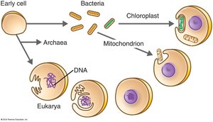
Endosymbiotic Theory
The endosymbiotic theory proposes that eukaryotic cells evolved from prokaryotic cells living inside one another. Mitochondria and chloroplasts in eukaryotes resemble bacteria in structure and function.
Binomial Nomenclature and Taxonomic Hierarchy
Organisms are classified using binomial nomenclature (Genus species, e.g., Homo sapiens) and organized into a hierarchy: Domain, Kingdom, Phylum, Class, Order, Family, Genus, Species, Subspecies.
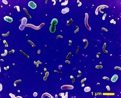
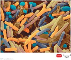
Microscopy and Preparation of Bacterial Specimens
Units of Measurement
1 cm = 10 mm
1 mm = 1000 μm (micrometers)
1 μm = 1000 nm (nanometers)
Types of Microscopes
Compound Light Microscope: Used for general laboratory observations.
Darkfield Microscope: Used to examine live, unstained microbes (e.g., spirochetes like Treponema pallidum).
Fluorescent Microscope: Uses UV light and fluorescent dyes to detect microbes in tissues or clinical specimens.
Phase-Contrast Microscope: Examines internal structures of living organisms without stains.
Electron Microscope (EM): Uses beams of electrons to view objects smaller than 0.2 μm (viruses, internal structures); includes TEM and SEM.
Preparation and Examination of Bacterial Specimens
Living State: Wet mount and hanging drop methods are used to observe motility.
Non-living State: Staining is used to emphasize features; heat fixing attaches bacteria to the slide and kills them.
Types of Stains
Simple Stain: Uses a single basic dye (e.g., crystal violet) to observe shape, size, and arrangement of cells.
Differential Stains:
Gram Stain: Differentiates bacteria based on cell wall composition. Gram-positive bacteria stain blue/purple (thick peptidoglycan), Gram-negative stain pink (thin peptidoglycan, thick lipid layer).
Acid-Fast Stain: Identifies bacteria with waxy cell walls (e.g., Mycobacterium); acid-fast positive cells stain red, negative stain blue.
Special Stains:
Flagella Stain: Visualizes number and arrangement of flagella.
Capsule Stain: Stains the background to visualize the capsule (e.g., Pasteurella multocida).
Endospore Stain: Identifies resistant, dormant structures (e.g., Clostridium, Bacillus).
Bioterrorism
Definition and Historical Examples
Bioterrorism is the deliberate or threatened use of bacteria, viruses, or toxins to cause disease, death, disruption, or fear. Historical examples include the use of plague-infected corpses, smallpox-contaminated blankets, and anthrax attacks.
1300s: Tartars catapulted plague-infected corpses, leading to the Black Death in Europe.
1700s: British soldiers gave smallpox-infected blankets to Native Americans.
World Wars: Use of anthrax and plague as biological weapons.
1984: Salmonella used in Oregon salad bars.
2001: U.S. Anthrax attacks.
Modern Bioterrorism and Public Health
The CDC categorizes biological agents by risk; those that are easily disseminated and cause high mortality are of greatest concern.
New technologies and vaccines are being developed to identify and prevent bioweapons attacks.
Aerosolized agents are considered the most likely method for future attacks due to ease of dissemination.
