Back
BackBiochemistry of the Genome & Mechanisms of Microbial Genetics: Core Concepts and Clinical Applications
Study Guide - Smart Notes
 Tailored notes based on your materials, expanded with key definitions, examples, and context.
Biochemistry of the Genome
DNA Nucleotides (Deoxyribonucleotides)
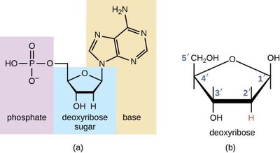
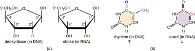
DNA is composed of building blocks called deoxyribonucleotides, each consisting of a deoxyribose sugar, a phosphate group, and a nitrogenous base. The five carbons in deoxyribose are numbered 1' through 5', which is important for understanding DNA structure and replication.
Deoxyribose sugar: A five-carbon sugar lacking an oxygen atom at the 2' position.
Phosphate group: Attached to the 5' carbon of the sugar.
Nitrogenous base: Attached to the 1' carbon; can be a purine or pyrimidine.
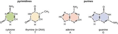
Pyrimidines and Purines
Nitrogenous bases in DNA are classified as purines (adenine and guanine, double-ringed) or pyrimidines (cytosine and thymine, single-ringed). Thymine is unique to DNA.
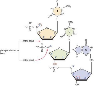
Sugar-Phosphate Backbone
Nucleotides are joined by phosphodiester bonds between the 5' phosphate of one nucleotide and the 3' hydroxyl of the next, forming the sugar-phosphate backbone of DNA. This backbone provides structural stability and directionality (5' to 3').
The Double Helix
Watson and Crick, building on Rosalind Franklin's work, proposed the double helix model of DNA. The two strands are antiparallel, with sugar-phosphate backbones on the outside and nitrogenous bases forming the rungs. The 5' and 3' ends indicate the directionality of each strand.
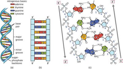
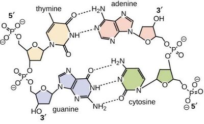
Complementary Base Pairing
Hydrogen bonds form between specific pairs of nitrogenous bases: adenine (A) pairs with thymine (T) via two hydrogen bonds, and guanine (G) pairs with cytosine (C) via three hydrogen bonds. This ensures accurate DNA replication and transcription.
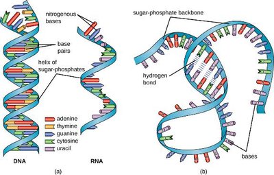
RNA Nucleotides (Ribonucleotides)
RNA is composed of ribonucleotides, which contain ribose sugar (with a hydroxyl group at the 2' position) and the base uracil (U) instead of thymine.
RNA Structure and Types
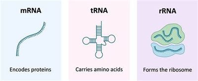
Unlike DNA, RNA is typically single-stranded but can fold into complex structures via intramolecular base pairing. There are three main types of RNA:
mRNA (messenger RNA): Carries genetic information from DNA to ribosomes.
tRNA (transfer RNA): Brings amino acids to the ribosome during translation.
rRNA (ribosomal RNA): Structural and catalytic component of ribosomes.
Central Dogma & Gene Expression
The Central Dogma
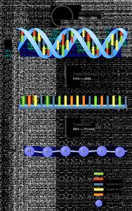
The central dogma of molecular biology describes the flow of genetic information: DNA is transcribed into mRNA, which is then translated into protein. This process is fundamental to all living organisms.
Gene Expression and Phenotype
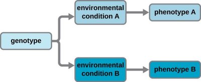
Gene expression determines phenotype. Although cells may share the same genotype, environmental conditions can lead to different patterns of gene expression, resulting in diverse phenotypes.
DNA Replication
Overview of DNA Replication
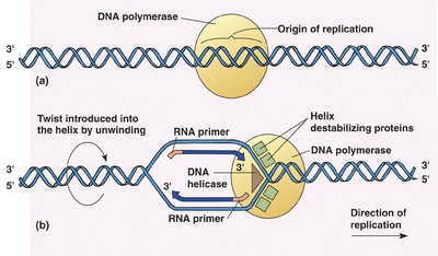
DNA replication is the process by which a cell copies its DNA before cell division (vertical gene transfer). Key enzymes include:
DNA helicase: Unwinds the double helix.
DNA primase: Synthesizes RNA primers.
DNA polymerase: Adds nucleotides to the 3' end of the new strand.
DNA ligase: Joins Okazaki fragments on the lagging strand.
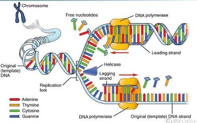
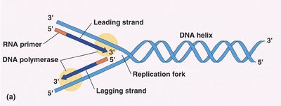
Initiation and Elongation
Replication begins at the origin of replication. Helicase unwinds DNA, and primase lays down RNA primers. DNA polymerase synthesizes new DNA in the 5' to 3' direction. The leading strand is synthesized continuously, while the lagging strand is synthesized discontinuously in Okazaki fragments.
Transcription and Translation
Transcription
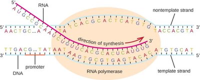
During transcription, RNA polymerase binds to a promoter sequence and synthesizes mRNA from the DNA template in the 5' to 3' direction. Transcription ends at a termination sequence.
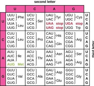
The Genetic Code
The genetic code is the relationship between mRNA codons (triplets of bases) and amino acids. It is universal and redundant. Start codon (AUG) codes for methionine; stop codons (UAA, UAG, UGA) terminate translation.
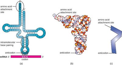
tRNA Structure and Function
tRNA molecules have an anticodon region that pairs with mRNA codons and an amino acid attachment site. They "translate" the nucleotide sequence into a polypeptide sequence.
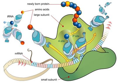
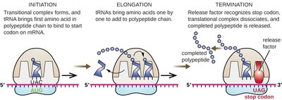
Translation
Translation occurs in the ribosome and involves three stages:
Initiation: tRNA binds to the start codon on mRNA, and the ribosomal subunits assemble.
Elongation: tRNAs bring amino acids to the ribosome, forming a growing polypeptide chain.
Termination: A stop codon is reached, and the completed polypeptide is released.
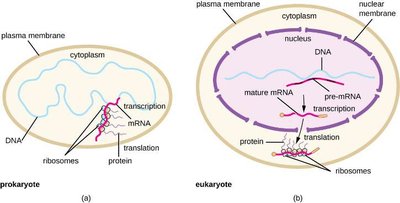
Prokaryotes vs. Eukaryotes: Gene Expression
In prokaryotes, transcription and translation occur simultaneously in the cytoplasm. In eukaryotes, transcription occurs in the nucleus and translation in the cytoplasm, with additional RNA processing steps.
Mutations
Types of Mutations
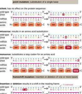
Mutations are changes in DNA sequence that can arise spontaneously or be induced. Types include:
Point mutation: Substitution of a single base.
Frameshift mutation: Insertion or deletion of bases, altering the reading frame.
Silent mutation: No effect on protein sequence.
Missense mutation: Changes one amino acid.
Nonsense mutation: Introduces a stop codon.
Clinical Focus: E. coli and Horizontal Gene Transfer
Pathogenic E. coli and Traveler's Diarrhea
Escherichia coli is a Gram-negative rod commonly found in the colon. While most strains are harmless, some are pathogenic and cause diarrheal diseases, especially after travel. Diagnosis involves culturing and PCR to detect virulence genes. Antibiotic therapy is usually avoided due to the risk of endotoxic shock.
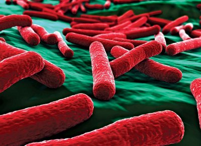
Pathogenic Strains of E. coli
ETEC (Enterotoxigenic E. coli): Causes traveler's diarrhea; produces heat-stable and heat-labile toxins.
EIEC (Enteroinvasive E. coli): Causes inflammatory disease of the large intestine.
EPEC (Enteropathogenic E. coli): Causes infantile diarrhea; forms pathogenicity islands.
EHEC (Enterohemorrhagic E. coli): Produces Shiga-toxin; can cause bloody diarrhea and HUS.
Horizontal Gene Transfer in Bacteria
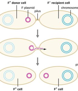
Bacteria can acquire new genetic traits via horizontal gene transfer:
Transformation: Uptake of DNA from the environment.
Transduction: Transfer of DNA by bacteriophages.
Conjugation: Direct transfer of DNA between cells via a pilus.
Streptococcus pyogenes and Skin Infections
S. pyogenes Skin Conditions
Streptococcus pyogenes is a Gram-positive coccus that can cause various skin infections, including cellulitis, erysipelas, and erythema nodosum. Its virulence factors include hyaluronidase, streptokinase, streptolysins, capsule, and M proteins.
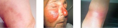
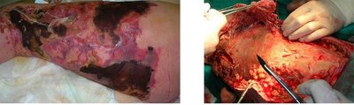
Necrotizing Fasciitis
Necrotizing fasciitis is a severe soft tissue infection, often caused by S. pyogenes, that can rapidly destroy tissue and requires aggressive treatment. Other causative agents include Klebsiella, Clostridium, E. coli, S. aureus, and Aeromonas.