 Back
BackBrock Biology of Microorganisms: The Microbial World (Chapter 1) - Study Notes
Study Guide - Smart Notes
 Tailored notes based on your materials, expanded with key definitions, examples, and context.
The Microbial World
Introduction to Microorganisms
Microorganisms, or microbes, are life forms too small to be seen by the human eye and require a microscope for observation. They are classified into prokaryotic (before nucleus), eukaryotic (true nucleus), and viruses (not free-living cells, require a host for reproduction).
Prokaryotic microbes: Include Bacteria (e.g., Streptococcus pyogenes), Cyanobacteria (e.g., Anabeana), and Archaea (e.g., Methanocaldococcus jannaschii).
Eukaryotic microbes: Include fungi (e.g., Candida albicans, Rhizopus stolonifer), protists (e.g., Trypansoma cruzi), animals (e.g., Trichuris vulpis), and plants (e.g., Spirogyra).
Viruses: Non-cellular entities composed of DNA or RNA surrounded by a protein coat, sometimes with an envelope. They require host cells to replicate.
Microorganisms: Importance and Applications
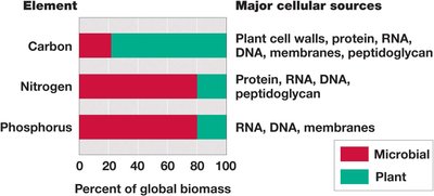
Microorganisms are the oldest form of life, constitute a major fraction of Earth's biomass, and have profound effects on human life, including infectious diseases, food and water safety, soil fertility, animal health, and fuel production. Pathogens are organisms that cause diseases.

Studying Microbes: Tools and Techniques
Microscopy and Culture Media
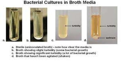
Microbes are studied using microscopes (compound light and electron microscopes) and culture media. Culture media can be liquid (broth), solid (agar), or slanted. Growth refers to an increase in cell number due to cell division, and a colony is a visible mass containing millions or billions of cells.
Plated media: Trypticase Soy Agar (TSA), Mannitol Salt Agar (MSA)
Liquid media: Trypticase Soy Broth (TSB), Glucose purple broth
Slanted media: TSA slants, Citrate Agar Slants
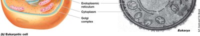
Structure and Activities of Microbial Cells
Basic Cell Structure
All cells share common structural elements:
Cytoplasmic (cell) membrane: Phospholipid bilayer that separates the cytoplasm from the external environment.
Cytoplasm: Aqueous mixture of macromolecules, small organics, ions, and ribosomes.
Ribosomes: Protein-synthesizing structures; bacteria have 70S ribosomes, eukaryotes have 80S ribosomes.
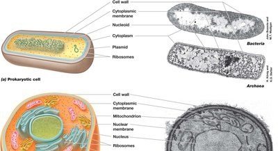
Cell Walls
Bacteria: Peptidoglycan (murein)
Acid-fast bacteria: Mycolic acids in cell walls
Archaea: Pseudopeptidoglycan (pseudomurein)
Fungi: Chitin
Plants: Cellulose
Animal cells: No cell walls, only cell membranes; sensitive to osmotic pressure
Mycoplasma: Bacteria without cell walls, pleomorphic, causes walking pneumonia
Genetic Material and Genome Organization
Genome: Full set of genes in a cell
Eukaryotic DNA: Linear chromosomes within a nucleus, large genome
Prokaryotic DNA: Single circular chromosome in nucleoid region, may have plasmids (antibiotic resistance), small and compact genome
Cell Activities
Metabolism: Chemical transformation of nutrients; includes aerobic, anaerobic, and facultative anaerobic processes
Enzymes: Protein catalysts for biochemical reactions
Transcription: DNA to RNA
Translation: RNA to protein
DNA replication: Copying the genome
Motility: Movement via flagella (bacteria) or cilia/flagella (eukaryotes)
Differentiation: Formation of specialized cells (endospores in bacteria, spores in fungi, pili for conjugation)
Intercellular communication: Chemical signaling (quorum sensing)
Evolution: Genetic changes passed to offspring
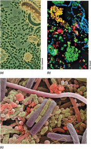
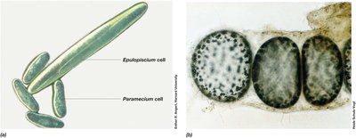
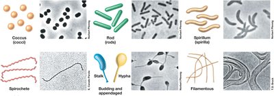
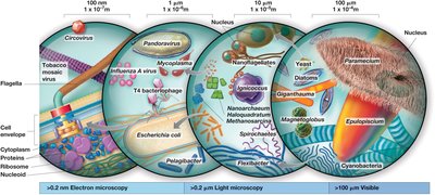
Cell Size and Morphology
Cell Size Range
Cell size and shape (morphology) vary widely among microbes. Prokaryotes range from 0.2 µm to 600+ µm in diameter, most between 0.5 and 10 µm. Eukaryotic cells are typically 5 to 100 µm in length.
Organism | Size (µm) | Morphology | Characteristics |
|---|---|---|---|
Thiomargarita namibiensis | 750 | Cocci in chains | Sulfur chemolithotroph |
Epulopiscium fishelsonia | 80 × 600 | Rods with tapered ends | Chemoorganotroph |
Escherichia coli | 1 × 2 | Rods | Chemoorganotroph |
Mycoplasma pneumoniae | 0.2 | Pleomorphic | Pathogenic bacterium |
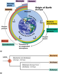
Introduction to Microbial Life
Three Domains of Life
All cellular life is classified into three domains: Bacteria, Archaea, and Eukarya.
Bacteria: Prokaryotes, usually undifferentiated single cells, 0.5–10 μm long, e.g., E. coli (rod-shaped), Staphylococcus aureus (cocci-shaped)
Archaea: Prokaryotes, often extremophiles, no known parasites or pathogens of plants and animals
Eukarya: Includes plants, animals, fungi, protists; first were unicellular, may have appeared two billion years ago; endosymbiotic theory explains origin of mitochondria and chloroplasts
Viruses
Obligate parasites, replicate only within host cells
Not cells, do not carry out metabolism
Small genomes of double- or single-stranded DNA or RNA
Classified by structure, genome composition, and host specificity
Microorganisms and the Biosphere
History of Life on Earth
Earth is 4.6 billion years old. First cells appeared between 3.8 and 4.3 billion years ago. The atmosphere was anoxic until ~2.6 billion years ago, supporting only anaerobic metabolisms. Cyanobacteria (oxygenic phototrophs) appeared ~2.6 billion years ago, and plants and animals ~0.5 billion years ago.
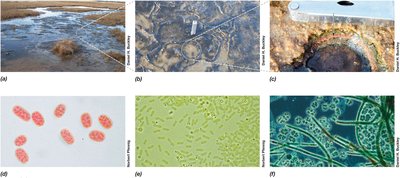
Microbial Ecology and Extremophiles
Microbial ecology studies how microbes affect animals, plants, and ecosystems. Extremophiles live in habitats too harsh for other life forms, such as hot springs, glaciers, high salt, acidity/alkalinity, and pressure.
Descriptive Term | Habitat | Domain | Genus, Species | Extreme Condition |
|---|---|---|---|---|
Hyperthermophile | Undersea hydrothermal vents | Archaea | Methanopyrus kandleri | High temperature (up to 122°C) |
Psychrophile | Sea ice | Bacteria | Psychromonas ingrahamii | Low temperature (-12°C) |
Acidophile | Acidic hot springs | Archaea | Picrophilus oshimae | Low pH (0.7) |
Halophile | Salterns | Archaea | Halobacterium salinarum | High salt (32% NaCl) |
Impact of Microorganisms on Human Society
Microorganisms as Disease Agents
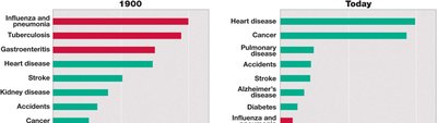
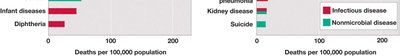
Microorganisms can be both beneficial and harmful. Pathogens cause disease, but most microbes are beneficial, contributing to vaccination, antibiotic therapy, water treatment, and food safety.
Microorganisms in Agriculture and Nutrition
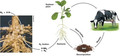
Nitrogen-fixing bacteria: Convert atmospheric nitrogen to ammonia for plant use
Cellulose-degrading microbes: In the rumen of cattle, help digest plant matter
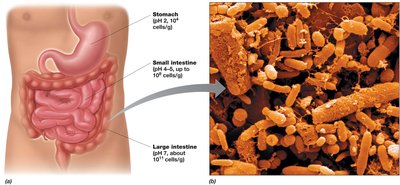
Gut microbiome: Digests complex carbohydrates in humans, synthesizes vitamins and nutrients
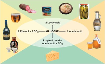
Microorganisms and Food
Negative impacts: Food spoilage and foodborne disease
Positive impacts: Food safety and preservation, production of dairy products (cheese, yogurt), fermented foods (sauerkraut, kimchi, pickles, chocolate, coffee, bread, alcohol)
Microorganisms and Industry
Industrial microbiology: Use of microbes in pharmaceuticals, brewing, and biotechnology
Biotechnology: Genetically engineered microbes produce high-value products
Biofuels: Production of methane and ethanol
Wastewater treatment and bioremediation: Cleaning up pollutants
Biofilms: Growth on submerged surfaces (pipes, drains, medical devices)


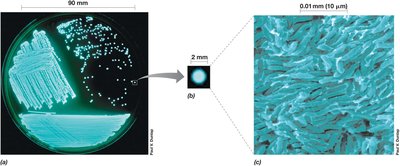
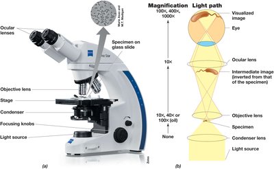
Microscopy and Discovery of Microorganisms
History of Microscopy
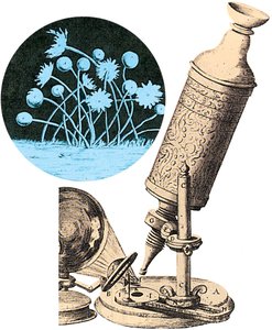
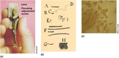
Microbiology began with the invention of the microscope. Robert Hooke first described microbes in 1665, and Antonie van Leeuwenhoek was the first to see bacteria.
Types of Light Microscopy
Bright-field: Visualizes specimens by differences in contrast
Phase-contrast: Amplifies differences in refractive index
Differential interference contrast: Enhances contrast in unstained cells
Dark-field: Light scattered by specimen, excellent for motility observation
Fluorescence: Visualizes specimens that fluoresce, widely used in diagnostics
Staining Techniques
Simple stains: Use basic dyes (methylene blue, crystal violet, safranin) to increase contrast
Negative stains: Stain the background, not the cell (Nigrosin, India ink, Congo Red)
Differential stains: Render different cells different colors; Gram stain distinguishes gram-positive (purple-violet) and gram-negative (red/pink) bacteria
Advanced Microscopy
Confocal scanning laser microscopy (CSLM): Generates three-dimensional images using a laser and computer
Electron microscopy: Uses electrons instead of light; includes transmission electron microscopes (TEM, see inside cells) and scanning electron microscopes (SEM, see cell surfaces)
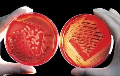
Microbial Cultivation and Historical Experiments
Aseptic Technique and Pure Cultures
Aseptic technique: Practices to maintain sterile media and solutions
Pure cultures: Cells from a single type of microorganism
Enrichment culture techniques: Isolate microbes with specific metabolic characteristics
Pasteur and Spontaneous Generation
Louis Pasteur disproved the theory of spontaneous generation using the swan-necked flask experiment, leading to sterilization methods and food preservation. He also developed vaccines for anthrax, fowl cholera, and rabies.
Koch and Infectious Disease
Robert Koch demonstrated the link between microbes and infectious diseases (germ theory), identified causative agents of anthrax, tuberculosis, and cholera, and developed Koch's postulates to link cause and effect in infectious disease. He also developed solid media for pure cultures.
Molecular Basis of Life and Evolution
Foundations of Molecular Biology
Rapid growth of bacteria under controlled conditions makes them excellent models for studying molecular biology, genetics, and biochemistry.
Genetic transfer in bacteria and the discovery that DNA is the genetic material were foundational (Griffith, Avery-MacLeod-McCarty, Watson, Crick, Franklin).
Woese and the Tree of Life
Ribosomal RNA (rRNA) sequencing enabled the construction of the phylogenetic tree of life, showing three domains: Bacteria, Archaea, and Eukarya.
LUCA (last universal common ancestor) is the root of the tree.
Most microbes have not been cultured yet; DNA sequencing technology allows study of entire genomes.
