 Back
BackCapsule Staining and Capsule-Forming Bacteria
Study Guide - Smart Notes
 Tailored notes based on your materials, expanded with key definitions, examples, and context.
Capsule Staining in Microbiology
Introduction to Bacterial Capsules
Many bacteria produce an extracellular capsule composed of polysaccharides or polypeptides. This structure enhances pathogenicity by protecting bacteria from host defenses and aiding in adherence to surfaces. Capsules are important virulence factors in several clinically significant bacteria.
Capsule: A gelatinous, non-ionic layer external to the cell wall, composed mainly of polysaccharides (sometimes polypeptides).
Function: Increases bacterial virulence, prevents phagocytosis, and assists in surface attachment.
Examples: Bacillus anthracis, Streptococcus pneumoniae, Streptococcus mutans (produces dextran), Klebsiella pneumoniae.
Principle of Capsule Staining
Capsule staining is a differential staining technique used to visualize bacterial capsules. Because capsules are non-ionic, they do not bind to most stains and appear as clear halos around stained cells against a colored background.
Stain Used: Carbol Fuchsin (a basic dye) stains the cell and background, but not the capsule.
Fixative: Albumin (egg white) is used to adhere cells to the slide and provide a background.
Result: Capsules appear as clear zones (halos) around pink/red-stained cells.
Capsule Staining Procedure
The following steps outline the capsule staining method, which allows visualization of the capsule as a clear halo around the bacterial cell.
Label the slide and add a scant loopful of distilled water (DiH2O).
Add a scant loopful of Mayer's Albumin.
Add a small amount of the bacterial organism (e.g., Klebsiella, Enterobacter, or Streptococcus) and mix well.
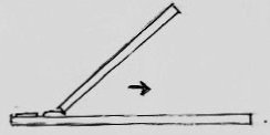
Spread the mixture across the slide using another slide held at a 45° angle to create a thin smear.
Allow the smear to air dry.
Gently heat-fix the slide by holding it at the tip of the burner flame for 5 seconds (until light smoke appears).
Cool the slide and flood with Ziehl-Neelsen Carbol Fuchsin for 10 seconds.
Wash gently with water and air-dry (do not blot).
Examine under oil immersion lens.
Examples of Capsule-Forming Bacteria
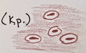
Klebsiella pneumoniae
Klebsiella pneumoniae is a Gram-negative, capsulated, straight rod-shaped bacterium. It is a facultative anaerobe with both respiratory and fermentative metabolism, commonly found in human feces, soil, water, and various foods. It is an opportunistic pathogen causing pneumonia, urinary tract infections (UTIs), and bacteremia.
Cell Morphology: 0.3–1.0 × 0.6–6 µm, occurs singly, in pairs, or short chains.
Capsule: Prominent, enhances virulence.
Optimal Temperature: ~37°C.
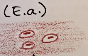
Enterobacter aerogenes
Enterobacter aerogenes is a Gram-negative, motile rod with peritrichous flagella. It is a facultative anaerobe and chemoorganotroph, found in water, soil, sewage, plants, and feces. It is an opportunistic pathogen.
Cell Morphology: 0.6–1.0 × 1.2–3.0 µm.
Motility: Peritrichous flagella.
Optimal Temperature: 30–37°C.
Streptococcus mutans
Streptococcus mutans is a Gram-positive, facultatively anaerobic coccus. It is a major contributor to dental caries (tooth decay) due to its ability to produce sticky dextran capsules, which facilitate adherence to tooth surfaces and biofilm formation.
Cell Morphology: Spherical (coccus), often in chains.
Capsule: Produces dextran, a sticky polysaccharide.
Habitat: Human oral cavity.

Summary Table: Capsule-Forming Bacteria
Bacterium | Gram Reaction | Shape & Arrangement | Capsule Composition | Pathogenicity |
|---|---|---|---|---|
Klebsiella pneumoniae | Negative | Rods, single/pairs/chains | Polysaccharide | Pneumonia, UTIs, bacteremia |
Enterobacter aerogenes | Negative | Rods, motile | Polysaccharide | Opportunistic infections |
Streptococcus mutans | Positive | Cocci, chains | Dextran (polysaccharide) | Dental caries |
Key Points
Capsule staining is essential for visualizing bacterial capsules, which are important virulence factors.
Capsules do not bind basic stains and appear as clear halos around stained cells.
Common capsule-forming bacteria include Klebsiella pneumoniae, Enterobacter aerogenes, and Streptococcus mutans.
