 Back
BackW7 Tues
Study Guide - Smart Notes
 Tailored notes based on your materials, expanded with key definitions, examples, and context.
Carbohydrate Catabolism & Fermentation
Metabolism Overview
Metabolism encompasses all chemical reactions occurring within living organisms, crucial for energy production and biosynthesis. These processes are mediated by enzymes, which are specialized proteins that catalyze biological reactions.
Enzymes: Proteins that accelerate biochemical reactions.
Endoenzymes: Function inside the cell.
Exoenzymes: Released from the cell to catalyze reactions outside.
Catabolism and Anabolism
Catabolism: Breakdown of large molecules (carbohydrates, lipids, proteins) into smaller units, releasing energy for cellular activities. Example: Glycolysis, digestion of food.
Anabolism: Synthesis of complex macromolecules from smaller units, requiring energy input. Example: Protein synthesis, muscle growth.
Carbohydrates: Structure and Classification
Carbohydrates are organic molecules with the general formula (CH2O)n, classified by size:
Monosaccharides: Simple sugars (e.g., glucose).
Oligosaccharides: 2–20 monosaccharide units.
Polysaccharides: 20 or more monosaccharide units (e.g., starch).
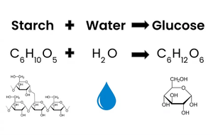
Starch Hydrolysis
Principle and Biochemical Basis
The starch hydrolysis test detects an organism's ability to produce the enzyme amylase, which breaks down starch into simpler sugars. Starch agar is a differential medium used to test for exoenzymes (hydrolytic enzymes) that degrade starch.
Hydrolysis: Breakdown of large substrates by addition of water.
Starch: Complex polysaccharide, too large to enter bacterial cells; exoenzymes degrade it into utilizable subunits.
Procedure
Divide starch agar plate into sectors and label.
Streak bacteria sample in a single line.
Incubate inverted at 35°C for 24 hours.
Flood plate with Gram’s iodine (indicator).
Examine for clear zone around bacterial growth.
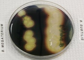
Interpretation of Results
Positive Test: Clear zone around growth after iodine addition indicates starch hydrolysis. Example: Bacillus species show strong amylase activity.
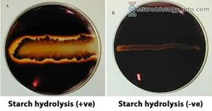
Negative Test: Dark blue coloration; starch remains intact. Example: Staphylococcus aureus, Staphylococcus epidermidis, E. coli.

Oxidative Fermentative (OF) Test
Principle and Media
The OF test differentiates bacteria based on their ability to metabolize glucose oxidatively (aerobic) or fermentatively (anaerobic and aerobic). OF-glucose deeps contain glucose, peptones, and bromothymol blue (pH indicator).
Oxidative bacteria: Produce acid only under aerobic conditions.
Fermentative bacteria: Produce acid under both aerobic and anaerobic conditions.
Non-saccharolytic bacteria: Do not metabolize glucose; may increase pH by amine production.
Procedure
Inoculate two tubes of OF-glucose medium.
Overlay one tube with mineral oil (anaerobic), leave the other open (aerobic).
Incubate at 35°C for 24–48 hours.
Observe growth, color change, and type of metabolism.
Results and Interpretation
Oxidative Result: Acid production in open tube (yellow), no change in oil-covered tube. Example: Pseudomonas aeruginosa, Acinetobacter species.
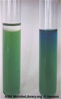
Fermentative Result: Acid production in both tubes (yellow); facultative anaerobes. Example: Enterobacteriaceae.
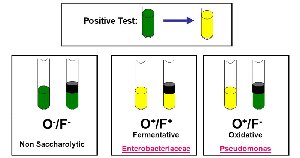
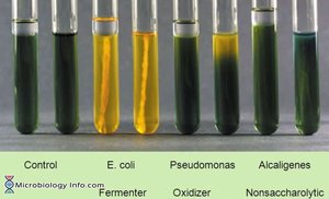
Negative Result: No color change or blue coloration (alkaline); non-saccharolytic. Example: Alcaligenes faecalis.
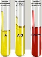
Carbohydrate Fermentation Tests
Principle and Media
Carbohydrate fermentation tests determine whether bacteria can ferment specific carbohydrates, producing acid and/or gas.
Basal medium: Contains a single carbohydrate (e.g., glucose, lactose, sucrose).
Phenol red: pH indicator (red = neutral, yellow = acidic).
Durham tube: Detects gas production (bubble formation).
Interpretation of Results
Fermenter with acid only: Yellow color, no gas.
Fermenter with acid and gas: Yellow color, gas bubble in Durham tube.
Non-fermenter: Red color, no acid or gas.
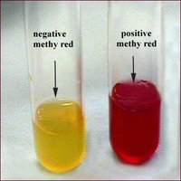
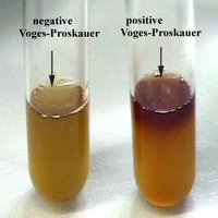
Methyl Red (MR) and Voges–Proskauer (VP) Tests
Principle and Procedures
The MR and VP tests differentiate Enterobacteriaceae and other Gram-negative rods based on glucose fermentation pathways.
MR Test: Detects strong acid production from mixed acid fermentation. Positive: Red color (pH < 4.4). Negative: Yellow/orange (pH > 6).
VP Test: Detects acetoin production (neutral fermentation). Positive: Red color after reagents added. Negative: No color change or copper discoloration.
Distinguishes: E. coli (MR+) from Enterobacter/Klebsiella (VP+).
Simmons Citrate Agar Test
Principle and Biochemical Basis
Simmons citrate agar is a defined medium used to determine if an organism can utilize citrate as its sole carbon source.
Citrate lyase: Hydrolyzes citrate to oxaloacetic acid and acetic acid.
CO2 production: Reacts with medium to form sodium carbonate, raising pH.
Bromthymol blue: pH indicator (green = neutral, blue = alkaline).
Interpretation of Results
Citrate positive: Growth and intense blue color (alkaline pH > 7.6). Example: Klebsiella, Enterobacter, Salmonella, most Proteus species.
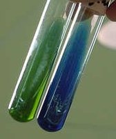
Citrate negative: No growth, medium remains green. Example: E. coli, Shigella.
Digestive System (Upper GI) Medical Terms
Key Terms
Esophago-: Esophagus
Gastro-: Stomach
Pyloro-: Pylorus (stomach outlet)
Cholecysto-: Gallbladder
Cholangio-: Bile ducts
Hepato-: Liver
Pancreato-: Pancreas
Emesis: Vomiting
Dysphagia: Difficulty swallowing
GERD: Gastroesophageal reflux disease (acid reflux)
Additional info: Academic context was added to clarify the principles, procedures, and interpretation of each biochemical test, as well as to provide definitions and examples for key terms and processes.
