 Back
BackCell Biology: Structure and Function of Prokaryotic and Eukaryotic Cells
Study Guide - Smart Notes
 Tailored notes based on your materials, expanded with key definitions, examples, and context.
Cell Biology: Prokaryotic and Eukaryotic Cells
Introduction to Cell Types
Cells are the fundamental units of life, and in microbiology, they are classified into two main types: prokaryotic and eukaryotic cells. Understanding their differences is essential for studying microbial structure, function, and diversity.
Prokaryotic Cells
General Characteristics
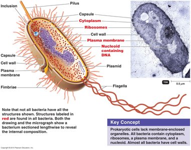
Prokaryotes ("pro" = before, "karyon" = nucleus) lack a membrane-bound nucleus.
DNA is found in a single, circular chromosome located in a region called the nucleoid.
They do not possess membrane-bound organelles (e.g., mitochondria).
All prokaryotes are single-celled organisms, including Bacteria and Archaea.
Bacterial Morphology (Shape)
Bacteria are classified by their shape, which is important for identification and understanding their function.
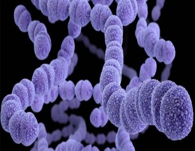
Coccus (plural: cocci): Spherical-shaped bacteria. Example: Streptococcus pyogenes.
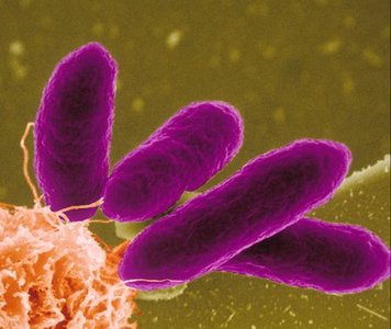
Bacillus (plural: bacilli): Rod-shaped bacteria. Example: Escherichia coli.
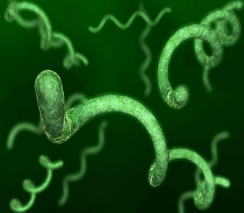
Spirillum (plural: spirilla): Spiral-shaped bacteria. Example: Treponema pallidum.
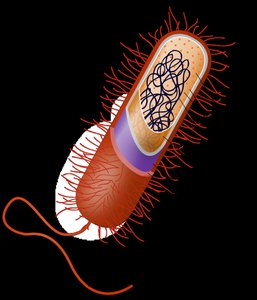
External Structures of Prokaryotes
Glycocalyx
A gelatinous, sticky polymer external to the cell wall, composed of polysaccharide, protein, or both.
When composed only of sugar, it is called an extracellular polysaccharide.
Secreted onto the outside of the cell wall.

Capsule vs. Slime Layer
Capsule: Firmly attached and organized glycocalyx; contributes to virulence by protecting against phagocytosis and aiding in adherence to surfaces.
Slime Layer: Loosely attached and unorganized glycocalyx.
Capsules are essential for the pathogenicity of some bacteria (e.g., Bacillus anthracis).

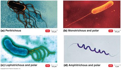
Flagella
Long, filamentous structures used for motility.
Composed of three parts: filament (flagellin protein), hook, and basal body (anchors flagellum to cell wall and membrane).
Flagellar Arrangements
Peritrichous: Flagella distributed over the entire cell surface.
Monotrichous: Single flagellum at one pole.
Lophotrichous: Two or more flagella at one or both ends.
Amphitrichous: Tuft of flagella at each end.
Bacterial Motility
Flagella rotate, allowing bacteria to move (run/swim or tumble).
Movement toward or away from stimuli is called taxis (e.g., chemotaxis for chemicals, phototaxis for light).
Pili and Fimbriae
Short, hair-like appendages found in many Gram-negative bacteria.
Fimbriae: Numerous, enable adherence to surfaces and colonization (e.g., Neisseria gonorrhoeae).
Pili: Longer, usually 1-2 per cell, involved in DNA transfer (conjugation).
Bacterial Cell Wall
The cell wall provides shape, protection, and is a key feature for bacterial classification.
Composed mainly of peptidoglycan, a polymer of sugars and amino acids.
Prevents cell rupture and protects against environmental changes.
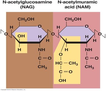
Peptidoglycan Structure
Consists of repeating disaccharide units: N-acetylglucosamine (NAG) and N-acetylmuramic acid (NAM).
Polysaccharide chains are cross-linked by short peptides, forming a strong lattice.
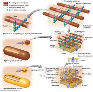
Gram-Positive vs. Gram-Negative Cell Walls
Feature | Gram-Positive | Gram-Negative |
|---|---|---|
Peptidoglycan Layer | Thick | Thin |
Teichoic Acids | Present | Absent |
Outer Membrane | Absent | Present (contains LPS) |
Lipopolysaccharide (LPS) | Absent | Present (endotoxin) |

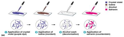
Gram Stain Procedure
Primary stain (crystal violet) stains all cells.
Iodine (mordant) forms a complex with crystal violet.
Alcohol wash decolorizes Gram-negative cells (outer membrane disrupted).
Counterstain (safranin) stains Gram-negative cells pink; Gram-positive remain purple.
Importance of Peptidoglycan
Unique to bacteria; target for antibiotics (e.g., penicillin) and host enzymes (e.g., lysozyme).
Disruption of peptidoglycan weakens the cell wall, leading to cell lysis.
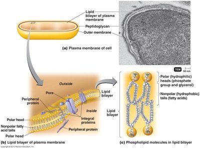
Plasma Membrane
Composed of a phospholipid bilayer with embedded proteins.
Acts as a semi-permeable barrier, controlling the movement of substances in and out of the cell.
Alcohol can disrupt the plasma membrane.
Cytoplasm
Gel-like substance inside the plasma membrane (~80% water).
Contains enzymes, nutrients, ribosomes, and genetic material.
Major structures: nucleoid, ribosomes, inclusion bodies, and sometimes endospores.
Nucleoid and Plasmids
Nucleoid: Region containing the bacterial chromosome (not membrane-bound).
Plasmids: Small, circular DNA molecules carrying non-essential genes (e.g., antibiotic resistance).
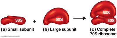
Ribosomes
Sites of protein synthesis, composed of rRNA and protein.
Prokaryotic ribosomes: 70S (50S large + 30S small subunit).
Target for antibiotics (e.g., streptomycin, erythromycin) due to structural differences from eukaryotic ribosomes (80S).
Inclusion Bodies
Storage granules for nutrients (e.g., sulfur, polysaccharides, lipids, enzymes).
Type and presence can aid in bacterial identification.

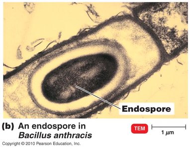
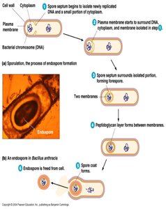
Endospores
Formed by some Gram-positive bacteria (e.g., Bacillus anthracis, Clostridium botulinum).
Highly resistant to heat, desiccation, chemicals, and radiation.
Allow bacteria to survive in harsh conditions; can remain dormant for long periods.
Eukaryotic Cells
General Characteristics
Contain a true, membrane-bound nucleus and multiple chromosomes.
Possess membrane-bound organelles (e.g., mitochondria, chloroplasts).
Can be unicellular (e.g., protozoa, some algae) or multicellular (e.g., fungi, plants, animals).

Examples of Eukaryotes
Protozoa: Unicellular organisms.
Fungi: Mostly multicellular (except yeasts).
Algae: Can be unicellular or multicellular.
Plants and Animals: Multicellular.
Eukaryotic Flagella and Cilia
Long, flexible structures containing protein and cytoplasm.
Move in a whip-like fashion (unlike the rotary motion of prokaryotic flagella).
Both are used for motility.
Eukaryotic Cell Wall
Absent in animal cells.
Structurally simpler than bacterial peptidoglycan.
Composed of cellulose (plants, algae) or chitin (fungi).
Eukaryotic Plasma Membrane
Similar in structure to prokaryotic plasma membrane but contains sterols (adds rigidity).
Capable of endocytosis (engulfing particles from outside the cell).
Eukaryotic Cytoplasm and Cytoskeleton
Located within the plasma membrane but outside the nuclear membrane.
Contains a complex cytoskeleton (protein filaments) for support, shape, and transport.
Membrane-Bound Organelles
Nucleus: Contains genetic material (DNA).
Mitochondria: Site of ATP synthesis ("powerhouse of the cell").
Chloroplasts: Site of photosynthesis (in plants and algae).

Additional info: This guide covers the essential structural and functional differences between prokaryotic and eukaryotic cells, with emphasis on features relevant to microbiology, such as cell wall composition, external structures, and mechanisms of motility and survival.
