 Back
Cell Structure and Function in Microbiology: Prokaryotes, Eukaryotes, and Viruses
Study Guide - Smart Notes
 Tailored notes based on your materials, expanded with key definitions, examples, and context.
Cell Structure and Function
Overview of Microbial Life Forms
Microorganisms are classified into three major groups: prokaryotes (Bacteria and Archaea), eukaryotes, and viruses. Each group exhibits unique structural and functional characteristics that distinguish them from one another and from multicellular life forms.
Prokaryotes: Simple, unicellular organisms lacking a nucleus and membrane-bound organelles. Includes Bacteria and Archaea.
Eukaryotes: More complex cells with a true nucleus and various internal organelles. Includes algae, protozoa, fungi, animals, and plants.
Viruses: Acellular entities that lack cellular structure and metabolism, requiring host cells for replication.
Characteristics of Life in Microbes
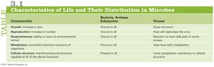
The table below summarizes the distribution of key life characteristics among Bacteria, Archaea, Eukaryotes, and Viruses.
Characteristic | Bacteria, Archaea, Eukaryotes | Viruses |
|---|---|---|
Growth | Occurs in all | Does not occur |
Reproduction | Occurs in all | Host cell replicates the virus |
Responsiveness | Occurs in all | Reaction to host cells seen in some viruses |
Metabolism | Occurs in all | Uses host cell metabolism |
Cellular structure | Present in all | Lacks cytoplasmic membrane or cellular structure |
Comparing Prokaryotic and Eukaryotic Cells
Prokaryotic Cell Structure
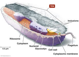
Prokaryotic cells are characterized by their simplicity and lack of internal compartmentalization. They are typically small (about 1.0 μm in diameter) and possess the following features:
No nucleus: DNA is located in a region called the nucleoid.
Lack of membrane-bound organelles: Cellular processes occur in the cytoplasm or at the cell membrane.
Cell wall: Provides structural support and shape.
External structures: May include glycocalyces, flagella, fimbriae, and pili.
Eukaryotic Cell Structure
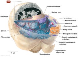
Eukaryotic cells are larger (10–100 μm in diameter) and more complex than prokaryotes. They contain:
True nucleus: DNA is enclosed within a nuclear envelope.
Membrane-bound organelles: Includes mitochondria, Golgi apparatus, endoplasmic reticulum, lysosomes, and more.
Complex cytoskeleton: Provides structural support and facilitates intracellular transport.
Endosymbiotic Theory
Origin of Eukaryotic Organelles
The endosymbiotic theory explains the origin of mitochondria and chloroplasts in eukaryotic cells. According to this theory:
Mitochondria and chloroplasts originated from free-living prokaryotes that were engulfed by ancestral eukaryotic cells.
These organelles contain their own circular DNA and 70S ribosomes, similar to bacteria.
They divide independently of the cell but rely on the host cell for many proteins.
Example: The mitochondria in animal cells and the chloroplasts in plant cells are believed to have evolved from aerobic and photosynthetic bacteria, respectively.
External Structures of Prokaryotic Cells
Glycocalyces
Glycocalyces are gelatinous, sticky substances that surround the outside of some prokaryotic cells. They are composed of polysaccharides, polypeptides, or both, and serve as protective and adhesive layers.
Capsule: Organized, firmly attached to the cell surface, and protects against desiccation and host immune responses (anti-phagocytic).
Slime layer: Loosely attached and less organized; aids in adherence to surfaces.
Flagella
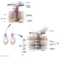
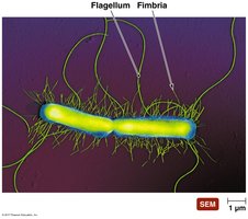
Flagella are long, whip-like appendages responsible for motility in many prokaryotes. They are composed of three main parts: filament, hook, and basal body.
Filament: Made of flagellin protein, forms a helical structure.
Hook: Connects the filament to the basal body.
Basal body: Anchors the flagellum to the cell wall and membrane, allowing rotation.
Arrangements of Flagella
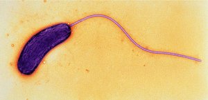
Monotrichous: Single flagellum at one end.
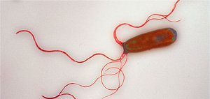
Lophotrichous: Tuft of flagella at one end.
Amphitrichous: One or more flagella at both ends.
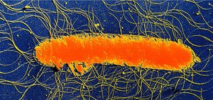
Peritrichous: Flagella distributed over the entire cell surface.
Function of Flagella
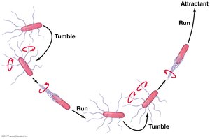
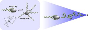
Flagella rotate to propel bacteria through their environment. Movement is powered by a proton motive force across the cell membrane. Bacteria exhibit taxis, moving toward or away from stimuli (e.g., chemotaxis, phototaxis, aerotaxis).
Runs: Straight movement, flagella rotate counterclockwise (CCW).
Tumbles: Random changes in direction, flagella rotate clockwise (CW).
Internal Flagella (Axial Filaments)
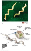
Some bacteria, such as spirochetes, possess internal flagella called axial filaments or endoflagella. These structures are located between the cell wall and outer membrane, enabling corkscrew-like movement.
Fimbriae and Pili
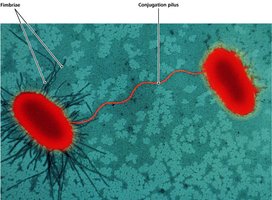
Fimbriae and pili are nonmotile, proteinaceous appendages found on many prokaryotes.
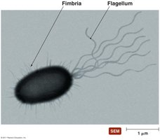
Fimbriae: Short, bristle-like projections used for attachment to surfaces, host tissues, and in biofilm formation. Hundreds may be present per cell.
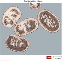
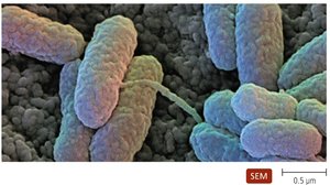
Pili (Conjugation Pili/Sex Pili): Longer than fimbriae but shorter than flagella. Usually one or two per cell, used for DNA transfer (conjugation) and specific attachment.
Prokaryotic Cell Walls
Structure and Function
The cell wall surrounds the cytoplasmic membrane and provides structural support, shape, and protection from osmotic lysis. It also serves as a barrier to certain antimicrobial substances. Animal cells lack cell walls, making bacterial cell walls a selective target for antibiotics.
Bacteria and Archaea differ in cell wall composition and structure.
Prokaryotic Cell Morphologies and Arrangements
Common Shapes
Coccus: Spherical
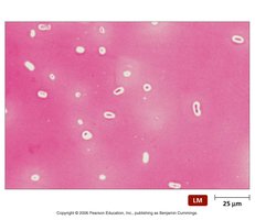
Bacillus: Rod-shaped
Spirillum: Rigid spiral
Spirochete: Flexible spiral
Vibrio: Comma-shaped
Coccobacillus: Short rod
Pleomorphic: Variable shape
Star-shaped: Rare, star-like appearance
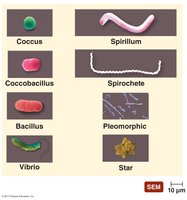
Spirillum vs. Spirochete
Spirilla: Rigid, thicker, external flagella, do not rotate as they move (e.g., Spirillum, Campylobacter).
Spirochetes: Flexible, thinner, internal endoflagella, rotate in corkscrew fashion (e.g., Leptospira, Borrelia, Treponema).
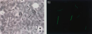
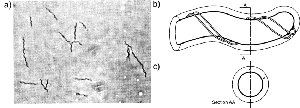
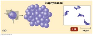
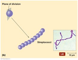
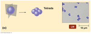
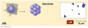
Arrangements of Cocci
Cocci can be arranged in various patterns depending on the plane of division and whether cells remain attached after division:
Diplococci: Pairs
Streptococci: Chains
Tetrads: Groups of four
Sarcinae: Cubes of eight
Staphylococci: Irregular clusters
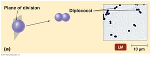
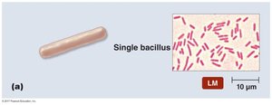
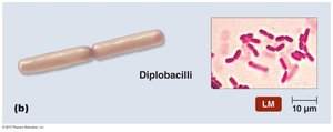
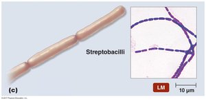
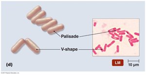
Arrangements of Bacilli
Bacilli may also form characteristic arrangements:
Single bacillus: Individual rods
Diplobacilli: Pairs
Streptobacilli: Chains
Palisade: Side-by-side arrangement
V-shape: Angled pairs
Additional info: Understanding the structure and function of prokaryotic and eukaryotic cells, as well as the unique features of viruses, is foundational for studying microbial physiology, genetics, and pathogenesis.
