 Back
BackCell Structure and Function in Microbiology
Study Guide - Smart Notes
 Tailored notes based on your materials, expanded with key definitions, examples, and context.
Cell Structure and Function
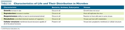
Characteristics of Life in Microbes
Microorganisms exhibit several fundamental characteristics that define life, including growth, reproduction, responsiveness, metabolism, and cellular structure. These traits are distributed differently among bacteria, archaea, eukaryotes, and viruses.
Growth: Increase in size; occurs in all cellular microbes but not in viruses.
Reproduction: Increase in number; occurs in all cellular microbes, while viruses replicate only within host cells.
Responsiveness: Ability to react to environmental stimuli; present in all cellular microbes, limited in viruses.
Metabolism: Controlled chemical reactions; present in all cellular microbes, absent in viruses.
Cellular Structure: Membrane-bound structure; present in all cellular microbes, lacking in viruses.
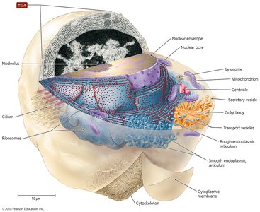
Types of Cells: Prokaryotic vs. Eukaryotic
Cells are classified as prokaryotic or eukaryotic based on their structural features. Prokaryotes include bacteria and archaea, while eukaryotes encompass algae, protozoa, fungi, animals, and plants.
Prokaryotes: Lack a nucleus, have a single circular chromosome, no organelles, and cell walls made of peptidoglycan (bacteria) or pseudomurein (archaea). Divide by binary fission.
Eukaryotes: Possess a nucleus, paired chromosomes, organelles, and polysaccharide cell walls (when present). Divide by mitosis.
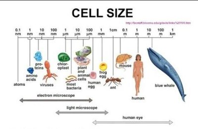
Cell Size Comparison
Cell sizes vary widely among different organisms, from tiny viruses to large eukaryotic cells and multicellular organisms. Most bacteria are much smaller than eukaryotic cells.
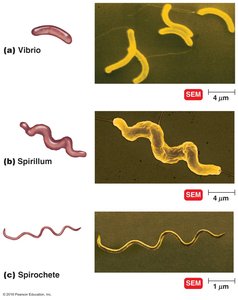
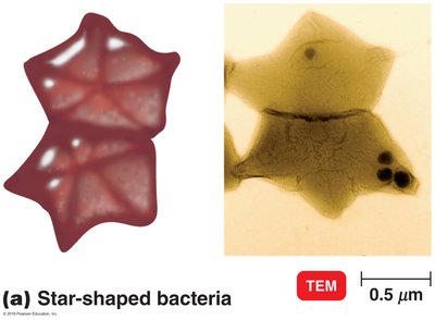
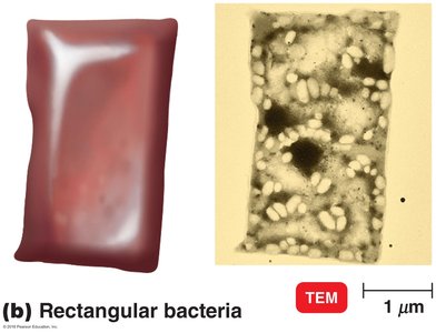
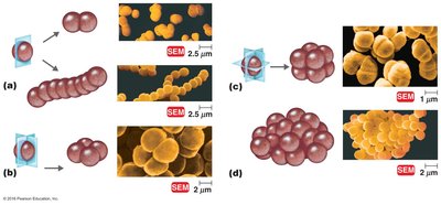
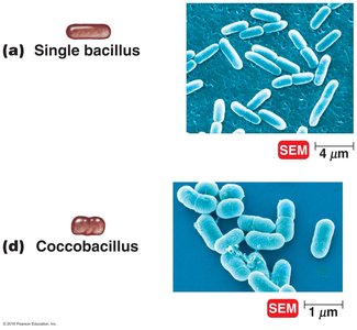
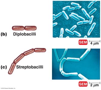
The Size, Shape, and Arrangement of Bacterial Cells
Bacterial Morphology
Bacteria display a variety of shapes and arrangements, which are important for identification and classification.
Shapes: Bacillus (rod-shaped), coccus (spherical), spiral (vibrio, spirillum, spirochete), star-shaped, rectangular.
Arrangements: Pairs (diplococci, diplobacilli), clusters (staphylococci), chains (streptococci, streptobacilli), tetrads, sarcinae (cubelike groups of eight).
Bacterial Cell Walls
Structure and Composition
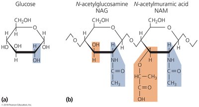
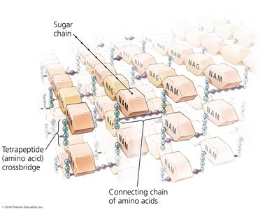
The cell wall provides structural support, shape, and protection from osmotic forces. It is a key target for antibiotics and is composed primarily of peptidoglycan.
Peptidoglycan: Polymer of repeating disaccharides (N-acetylglucosamine, NAG, and N-acetylmuramic acid, NAM) linked by polypeptides.
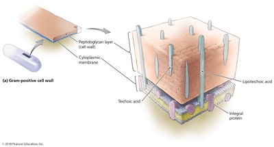
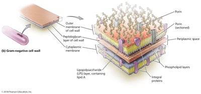
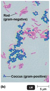
Gram-Positive vs. Gram-Negative Cell Walls
Bacterial cell walls are classified as Gram-positive or Gram-negative based on their structure and response to Gram staining.
Gram-Positive: Thick peptidoglycan layer, teichoic acids, lipoteichoic acids, purple after Gram stain.
Gram-Negative: Thin peptidoglycan layer, outer membrane with lipopolysaccharide (LPS), pink after Gram stain. Lipid A in LPS can cause fever and shock.
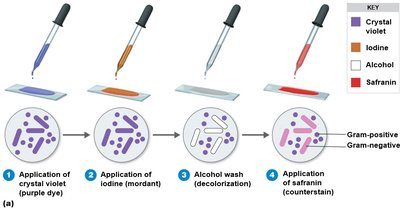
Gram Staining Procedure
Gram staining differentiates bacteria based on cell wall properties. The process involves application of crystal violet, iodine, alcohol wash, and safranin.
Gram-positive: Retain crystal violet, appear purple.
Gram-negative: Lose crystal violet, take up safranin, appear pink.
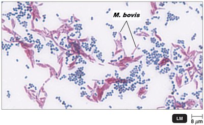
Special Cell Walls: Acid-Fast and Atypical
Some bacteria have atypical cell walls, such as acid-fast bacteria (e.g., Mycobacterium), which contain mycolic acid, and mycoplasmas, which lack cell walls.
Acid-fast: Waxy lipid (mycolic acid) bound to peptidoglycan; stain red with carbolfuchsin.
Mycoplasmas: Lack cell walls, have sterols in plasma membrane.
Archaea: May lack cell walls or have walls of pseudomurein.
Structures External to the Cell Wall
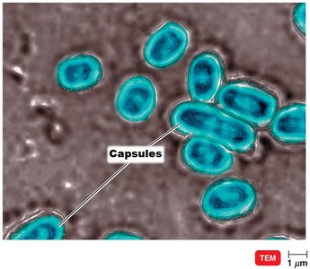
Glycocalyx
The glycocalyx is a gelatinous, sticky substance surrounding the outside of the cell, composed of polysaccharides and/or polypeptides. It exists as either a capsule (organized, firmly attached) or a slime layer (unorganized, loosely attached).
Capsule: Prevents phagocytosis, contributes to virulence.
Slime layer: Facilitates attachment to surfaces and biofilm formation.
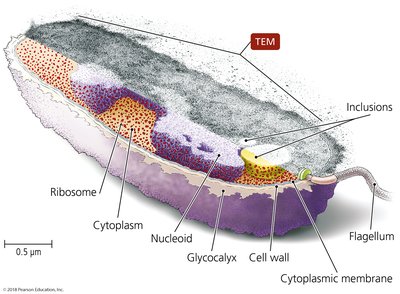
Flagella
Flagella are long, whip-like structures responsible for bacterial motility. They consist of a filament, hook, and basal body, and can be arranged in various patterns (monotrichous, lophotrichous, amphitrichous, peritrichous).
Function: Movement toward or away from stimuli (taxis), rotation for "run" or "tumble".
Fimbriae and Pili
Fimbriae are short, bristlelike projections used for adhesion and biofilm formation. Pili are longer, specialized fimbriae involved in motility and DNA transfer (conjugation).
Fimbriae: Important for attachment and biofilm formation.
Pili: Facilitate DNA transfer between cells.
Bacterial Cytoplasmic Membranes
Structure and Function
The cytoplasmic membrane is a phospholipid bilayer with embedded proteins, described by the fluid mosaic model. It is selectively permeable and involved in energy storage, ATP production, and maintaining gradients.
Integral and peripheral proteins: Facilitate transport and other functions.
Chromatophores: Membrane foldings with photosynthetic pigments in some bacteria.
Transport Across Membranes
Substances move across membranes by passive (diffusion, facilitated diffusion, osmosis) or active (active transport, group translocation) processes.
Passive: No energy required; moves substances from high to low concentration.
Active: Requires energy (ATP or PEP); moves substances against concentration gradient.
Cytoplasm of Bacteria
Cytosol and Inclusions
The cytosol is the liquid portion of the cytoplasm, containing water and dissolved substances. Inclusions are reserve deposits of chemicals such as phosphate, polysaccharides, lipids, and gas vacuoles.
Nucleoid and Plasmids
The nucleoid contains the bacterial chromosome (circular DNA), while plasmids are extrachromosomal genetic elements carrying non-essential genes.
Ribosomes and Cytoskeleton
Ribosomes are the sites of protein synthesis, composed of protein and rRNA (70S in prokaryotes). The cytoskeleton provides structural support and aids in cell division and movement.
Endospores
Endospores are highly resistant structures formed by some bacteria (e.g., Bacillus, Clostridium) as a defense against unfavorable conditions. They can survive extreme heat, radiation, and chemicals.
The Evolution of Eukaryotes
Endosymbiotic Theory
The endosymbiotic theory proposes that eukaryotes evolved when larger bacterial cells engulfed smaller ones, which became organelles such as mitochondria and chloroplasts. This theory is supported by similarities in DNA and structure between these organelles and certain bacteria.
Characteristic | Bacteria, Archaea, Eukaryotes | Viruses |
|---|---|---|
Growth | Occurs in all | Growth does not occur |
Reproduction | Occurs in all | Host cell replicates virus |
Responsiveness | Occurs in all | Reacts to host cells in some viruses |
Metabolism | Controlled chemical reactions | Viruses lack cell metabolism |
Cellular structure | Present in all | Viruses lack cytoplasmic membrane or cellular structure |
Additional info: These notes expand on the original lecture slides by providing definitions, examples, and context for each major concept, ensuring a comprehensive and self-contained study guide for microbiology students.
