 Back
BackCell Structure and Function: Chapter 3
Study Guide - Smart Notes
 Tailored notes based on your materials, expanded with key definitions, examples, and context.
Cell Structure and Function
Introduction
This chapter explores the fundamental structures and functions of microbial cells, focusing on the differences and similarities among prokaryotes, eukaryotes, and viruses. Understanding these cellular features is essential for comprehending microbial physiology, pathogenesis, and the basis for many clinical interventions.
Characteristics of Life in Microbes
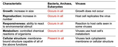
Defining Life Processes
Growth: Increase in size; occurs in all cellular life forms but not in viruses.
Reproduction: Increase in number; all cells reproduce, but viruses rely on host cells.
Responsiveness: Ability to react to environmental stimuli; present in all cells, some viruses show limited responsiveness.
Metabolism: Controlled chemical reactions; occurs in all cells, viruses use host metabolism.
Cellular Structure: Membrane-bound structure capable of all above functions; present in all cells, absent in viruses.
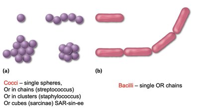
Types of Cells: Prokaryotes vs. Eukaryotes
Overview and Examples
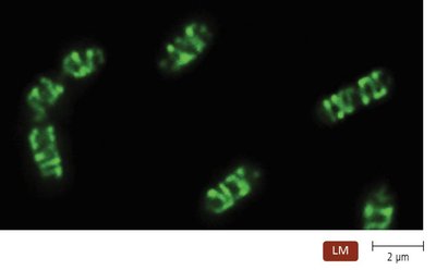
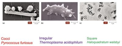
Prokaryotes: Lack a nucleus and membrane-bound organelles; include Bacteria and Archaea. Typically 1.0 μm or smaller.
Eukaryotes: Have a nucleus and internal membrane-bound organelles; include algae, protozoa, fungi, animals, and plants. Typically 10–100 μm.
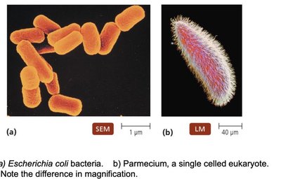
Example: Escherichia coli (prokaryote) vs. Paramecium (eukaryote).
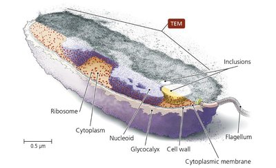
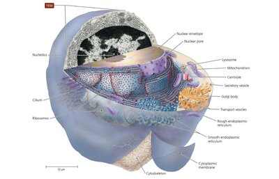
Cell Structure Diagrams
Typical prokaryotic and eukaryotic cells have distinct internal and external features.
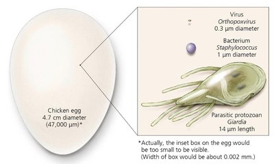
Cell Size Comparison
Cells and viruses vary greatly in size, from large eukaryotic cells to tiny viruses.
External Structures of Bacterial Cells
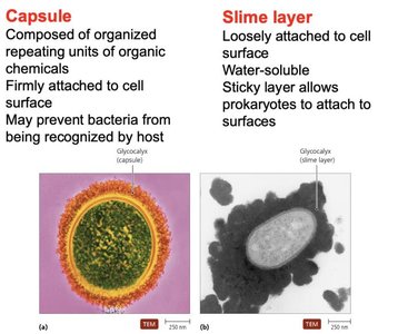
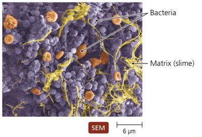
Glycocalyx
The glycocalyx is a gelatinous, sticky substance surrounding the outside of some bacterial cells, composed of polysaccharides, polypeptides, or both. It provides protection and aids in attachment.
Capsule: Organized, firmly attached, may prevent recognition by host immune system.
Slime Layer: Loosely attached, water-soluble, helps bacteria adhere to surfaces.
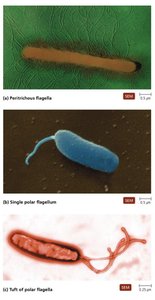
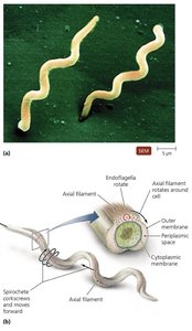
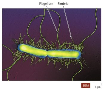
Flagella
Flagella are long, whip-like structures responsible for bacterial motility. They consist of a filament, hook, and basal body, and can be arranged in various patterns (peritrichous, polar, tufts).
Enable movement via rotation (runs and tumbles).
Allow taxis: movement toward or away from stimuli.
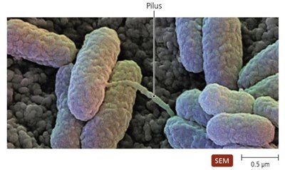
Fimbriae and Pili
Fimbriae: Short, bristle-like projections for adhesion to surfaces and other cells; important in biofilm formation.
Pili: Longer than fimbriae, usually one or two per cell; involved in DNA transfer (conjugation).
Bacterial Cell Walls
Structure and Function
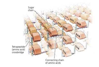
Provide shape, structural support, and protection from osmotic pressure.
Composed primarily of peptidoglycan (a mesh-like polymer of sugars and amino acids).
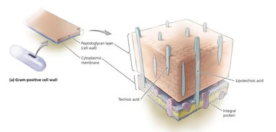
Gram-Positive vs. Gram-Negative Cell Walls
Gram-Positive: Thick peptidoglycan layer, teichoic acids, stains purple, may contain mycolic acid (acid-fast bacteria).
Gram-Negative: Thin peptidoglycan, outer membrane with lipopolysaccharide (LPS), stains pink, lipid A (endotoxin) can trigger strong immune responses.

Bacteria Without Cell Walls
Some bacteria lack cell walls and are often mistaken for viruses due to their small size.
They retain other prokaryotic features such as ribosomes.
Bacterial Cytoplasmic Membranes
Structure
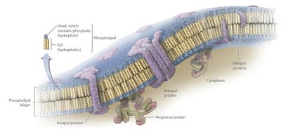
Composed of a phospholipid bilayer with embedded proteins (integral and peripheral).
Described by the fluid mosaic model.

Function
Controls passage of substances (selectively permeable).
Maintains concentration and electrical gradients.
In photosynthetic bacteria, harvests light energy.
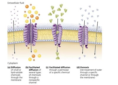
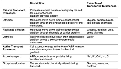
Transport Processes
Passive: Diffusion, facilitated diffusion, osmosis (no energy required).
Active: Active transport, group translocation (require energy, often ATP).
Cytoplasm of Bacteria
Components
Cytosol: Liquid portion, mostly water, contains DNA in the nucleoid region.
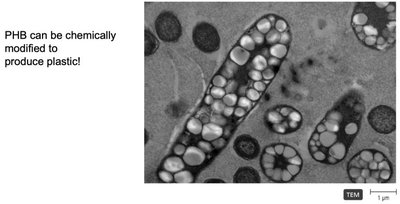
Inclusions: Reserve deposits of chemicals (e.g., PHB granules).
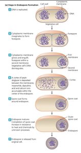
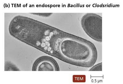
Endospores: Highly resistant structures formed under stress; allow survival in extreme conditions.
Cytoplasm of Eukaryotes
Nonmembranous Organelles
Ribosomes: Sites of protein synthesis; 80S in eukaryotes (60S + 40S subunits), 70S in prokaryotes.
Cytoskeleton: Network of fibers (microtubules, microfilaments, intermediate filaments) for shape, support, and movement.
Centrioles and Centrosome: Involved in cell division and organization of microtubules (not in all eukaryotes).
Membranous Organelles
Nucleus: Contains DNA, nucleolus (site of rRNA synthesis), surrounded by nuclear envelope with pores.
Endoplasmic Reticulum (ER): RER (with ribosomes) synthesizes proteins; SER synthesizes lipids and detoxifies.
Golgi Body: Processes and packages molecules for export.
Mitochondria: Site of ATP production; contains its own DNA and 70S ribosomes.
Chloroplasts: Found in photosynthetic eukaryotes; site of photosynthesis, contains DNA and 70S ribosomes.
External Structures of Archaea
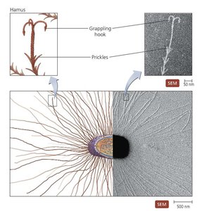
Glycocalyces, Flagella, Fimbriae, and Hami
Glycocalyces help in biofilm formation and adhesion.
Flagella are structurally different from bacterial flagella.
Fimbriae and unique hami (grappling hook-like structures) aid in attachment.
Cell Walls and Cytoplasmic Membranes
Most archaea have cell walls (no peptidoglycan), composed of specialized polysaccharides and proteins.
All have cytoplasmic membranes for maintaining gradients and transport.
Cytoplasm of Archaea
Similar to bacteria: 70S ribosomes, fibrous cytoskeleton, circular DNA.
Differences: unique ribosomal proteins, metabolic enzymes, and genetic code more similar to eukaryotes.
External Structure of Eukaryotic Cells
Glycocalyces and Cell Walls
Glycocalyces are less organized than in prokaryotes; aid in cell recognition, adhesion, and protection.
Cell walls (in fungi, algae, plants, some protozoa) are composed of polysaccharides (cellulose, chitin, glucomannan).
Cytoplasmic Membranes
Fluid mosaic of phospholipids and proteins; contain sterols for fluidity.
Membrane rafts organize proteins and lipids for signaling and transport.
Control movement into and out of the cell.
Endocytosis and Exocytosis
Active transport processes unique to eukaryotes for importing (endocytosis) and exporting (exocytosis) large molecules.
Flagella and Cilia
Eukaryotic flagella are structurally distinct from prokaryotic flagella; composed of microtubules, move by undulation, not rotation.
Cilia are shorter, more numerous, and move substances past the cell surface or propel the cell.
Clinical Application: E. coli Infection
Case Study Summary
Background: Patient with kidney infection due to E. coli (Gram-negative bacterium).
Pathogenesis: Stopping antibiotics early allowed E. coli to spread, causing systemic inflammation.
Key Cellular Component: Lipid A (endotoxin) from LPS in Gram-negative cell wall triggers inflammation, fever, and shock.
Virulence Factors: Flagella (motility), fimbriae (adhesion), and pili (DNA transfer and adhesion) help E. coli establish infection and spread.
Summary Table: Comparison of Prokaryotic, Eukaryotic, and Archaeal Cells
Feature | Bacteria | Archaea | Eukaryotes |
|---|---|---|---|
Nucleus | No | No | Yes |
Cell Wall | Peptidoglycan (most) | Polysaccharides/proteins (no peptidoglycan) | Polysaccharides (some) |
Ribosomes | 70S | 70S (different proteins) | 80S (70S in organelles) |
Membrane-bound Organelles | No | No | Yes |
Flagella | Yes (simple) | Yes (distinct structure) | Yes (complex, microtubules) |
