 Back
BackCell Structure and Function in Microbiology
Study Guide - Smart Notes
 Tailored notes based on your materials, expanded with key definitions, examples, and context.
Cell Structure and Function
Overview of Prokaryotic and Eukaryotic Cells
Microbial cells are classified into two main types: prokaryotes and eukaryotes. Understanding their structural differences is fundamental in microbiology.
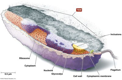
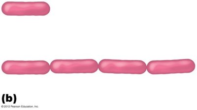
Prokaryotes: Lack a nucleus and membrane-bound organelles; include domains Bacteria and Archaea; typically small (~1.0 µm diameter); transcription and translation occur simultaneously.
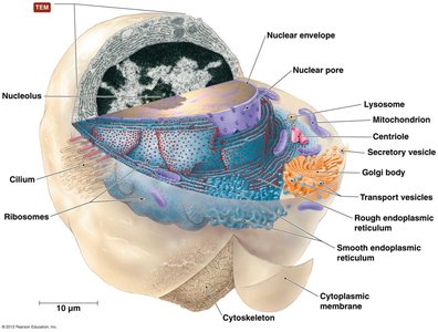
Eukaryotes: Possess a nucleus and internal membrane-bound organelles; larger (10–100 µm diameter); more complex; include algae, protozoa, fungi, animals, and plants.
Common Cell Structures
Both prokaryotic and eukaryotic cells share certain structural features:
Cell wall (CW)
Cell membrane (CM)
Cytoplasm
Glycocalyx
Flagella
Fimbriae
Pili
External Structures of Bacterial Cells
Glycocalyx: Slime Layer & Capsule
The glycocalyx is a gelatinous, sticky substance outside the cell, composed of polysaccharides, polypeptides, or both. It plays a crucial role in protection and attachment.
Slime layer: Loosely attached, water-soluble, aids in attachment to surfaces and formation of biofilms.
Capsule: Firmly attached, organized, prevents phagocytosis and desiccation, essential for pathogenicity.
Example: Streptococcus mutans forms a slime layer on teeth, leading to dental plaque and cavities.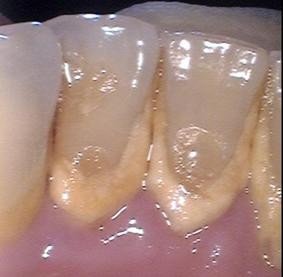
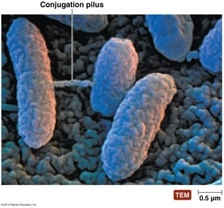
Fimbriae & Pili
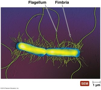
Fimbriae and pili are rodlike proteinaceous extensions used for attachment and genetic exchange.
Fimbriae: Sticky, bristle-like projections, shorter than flagella, important in biofilm formation.
Pili: Longer than fimbriae, hollow, non-motile; conjugation pili mediate DNA transfer between cells.
Biofilms
Biofilms are communities of bacteria embedded in a self-produced matrix, often involving glycocalyx and pili. They are highly resistant to antibiotics and play a major role in human infections and industrial problems.
Found on teeth (dental plaque), medical devices, and mucosal surfaces.
Can protect against pathogens (e.g., Lactobacilli biofilms in the vagina).
Flagella and Motility
Structure and Function of Flagella
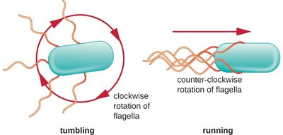
Flagella are long, whip-like appendages responsible for bacterial motility (taxis). They consist of a filament, hook, and basal body, and are made of flagellin protein.
Rotation propels bacteria; movement consists of "runs" (straight) and "tumbles" (direction change).
Movement is in response to stimuli: chemotaxis (chemical), phototaxis (light).
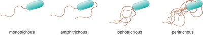
Flagellar Arrangements
Bacteria exhibit different flagellar arrangements:
Monotrichous: Single flagellum
Amphitrichous: Flagella at both ends
Lophotrichous: Tuft of flagella at one end
Peritrichous: Flagella all over the surface
Endoflagella in Spirochetes
Spirochetes possess endoflagella (axial filaments) located between the cell membrane and outer membrane, enabling corkscrew motility through viscous environments.
Example: Treponema pallidum (syphilis), Borrelia burgdorferi (Lyme disease)
Bacterial Cell Walls
Structure and Function
The bacterial cell wall provides structural support, shape, and protection from osmotic forces and antimicrobial drugs. It is primarily composed of peptidoglycan (repeating NAG & NAM units).
Two main types: Gram-positive and Gram-negative
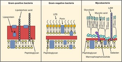
Gram-Positive Cell Walls
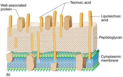
Gram-positive bacteria have thick peptidoglycan layers and unique chemicals like lipoteichoic acids, which anchor the cell wall and facilitate ion passage.
Acid-fast bacteria (e.g., Mycobacterium) contain mycolic acid for desiccation resistance.
Gram-Negative Cell Walls
Gram-negative bacteria have a thin peptidoglycan layer and an outer membrane containing lipopolysaccharide (LPS), which includes toxic Lipid A.
LPS acts as an endotoxin, triggering inflammation, fever, and shock when released.
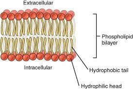
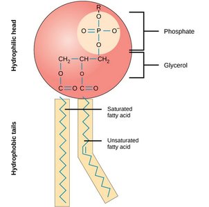
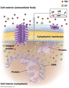
Bacterial Cell Membranes
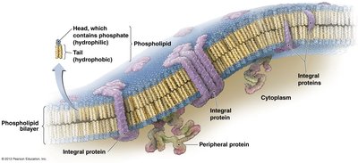
Phospholipid Bilayer Structure
The cell membrane is a phospholipid bilayer with embedded proteins, following the fluid mosaic model. It is selectively permeable and crucial for energy storage and transport.
Integral and peripheral proteins facilitate transport and communication.

Membrane Functions
Harvest light energy (photosynthetic bacteria)
Maintain concentration and electrical gradients
Selective permeability
Energy storage
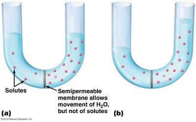
Transport Across Membranes
Transport mechanisms include passive (diffusion, facilitated diffusion, osmosis) and active (active transport, group translocation) processes.
Passive transport: No energy required
Active transport: Requires energy (ATP)
Group translocation: Substance chemically modified during transport
Cytoplasm of Bacteria
Inclusions
Bacterial cytoplasm contains inclusions—storage granules of lipids, starch, nitrates, phosphates, and sulfur. Polyhydroxybutyrates (PHB) are lipid polymers stored by stressed bacteria and can be used to produce biodegradable plastics.
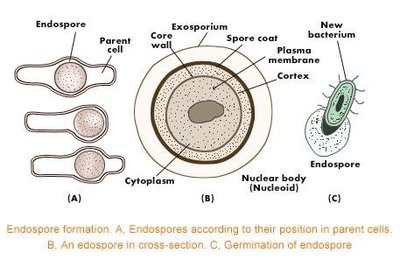
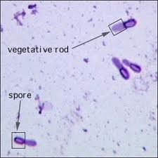
Endospores
Endospores are highly resistant, dormant structures formed by some Gram-positive bacteria (e.g., Bacillus, Clostridium) in response to unfavorable conditions. They are a major concern in food and healthcare industries due to their resistance to heat, drying, radiation, and chemicals.
Endospores contain DNA-binding proteins and are dehydrated for resistance.
Germinate when conditions become favorable.
Nonmembranous Organelles
Bacterial cytoplasm contains ribosomes (sites of protein synthesis) and a cytoskeleton (scaffolding protein for cell shape).
Summary Table: Comparison of Prokaryotic and Eukaryotic Cell Structures
Feature | Prokaryotes | Eukaryotes |
|---|---|---|
Nucleus | Absent | Present |
Membrane-bound organelles | Absent | Present |
Cell wall | Peptidoglycan (Bacteria), variable (Archaea) | Cellulose (plants), chitin (fungi), absent (animals) |
Size | ~1.0 µm | 10–100 µm |
Flagella | Simple, made of flagellin | Complex, made of microtubules |
DNA | Circular, in nucleoid | Linear, in nucleus |
