 Back
BackCell Structure and Function in Microbiology: Prokaryotic and Eukaryotic Cells
Study Guide - Smart Notes
 Tailored notes based on your materials, expanded with key definitions, examples, and context.
Processes of Life
Fundamental Characteristics of Living Cells
All living cells exhibit several essential processes that define life. These include growth, reproduction, responsiveness, and metabolism. Understanding these processes is foundational to microbiology.
Growth: Increase in size or number of cells.
Reproduction: Production of new cells or organisms.
Responsiveness: Ability to respond to environmental stimuli.
Metabolism: Sum of all chemical reactions within a cell.
Types of Cells
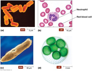
Examples of Cell Types
Cells can be classified into prokaryotic and eukaryotic types, each with distinct structural features and functions. The diversity of cell types is illustrated below.
Prokaryotic cells: Bacteria and archaea, lacking a nucleus.
Eukaryotic cells: Algae, protozoa, fungi, animals, and plants, possessing a nucleus and organelles.
Prokaryotic and Eukaryotic Cells: An Overview
Prokaryotes
Prokaryotic cells are characterized by their simple structure and lack of internal membrane-bound organelles. They are typically small and include bacteria and archaea.
Lack nucleus
Lack internal structures bound with phospholipid membranes
Small size: ~1.0 µm in diameter
Simple structure
Composed of: Bacteria and archaea
Eukaryotes
Eukaryotic cells are larger and more complex, containing a nucleus and various membrane-bound organelles. They are found in algae, protozoa, fungi, animals, and plants.
Have nucleus
Have internal membrane-bound organelles
Larger size: 10–100 µm in diameter
Complex structure
Composed of: Algae, protozoa, fungi, animals, plants
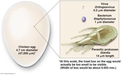
Cell Size Comparison
Approximate Size of Various Types of Cells
Microbial cells vary greatly in size, from viruses to protozoa, and even larger eukaryotic cells. Understanding these differences is important for microscopy and classification.
Virus: ~0.3 µm diameter
Bacterium: ~1 µm diameter
Parasitic protozoan: ~14 µm length
Chicken egg: ~4.7 cm diameter (47,000 µm)
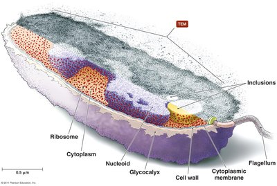
External Structures of Bacterial Cells
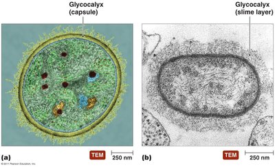
Glycocalyces
Bacterial cells often possess a glycocalyx, a gelatinous, sticky substance surrounding the cell. It is composed of polysaccharides, polypeptides, or both, and plays a role in protection and adherence.
Capsule: Organized, firmly attached, may prevent recognition by host.
Slime layer: Loosely attached, water soluble, aids in surface attachment.
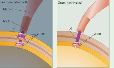
Flagella
Structure and Function
Flagella are long, whip-like structures responsible for bacterial motility. They consist of a filament, hook, and basal body, which anchors the flagellum to the cell wall and membrane.
Filament: Extends outward from the cell.
Hook: Connects filament to basal body.
Basal body: Anchors flagellum, composed of rods and rings.

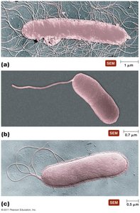
Flagella Arrangements
Bacteria exhibit various flagellar arrangements, which are important for classification and motility.
Monotrichous: Single flagellum
Lophotrichous: Tuft of flagella at one end
Amphitrichous: Flagella at both ends
Peritrichous: Flagella all over the surface
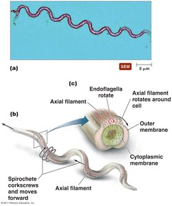
Axial Filaments (Spirochetes)
Some bacteria, such as spirochetes, possess axial filaments (endoflagella) that allow corkscrew-like movement.
Axial filament: Located between cell membrane and outer membrane
Function: Rotation causes cell to move in a corkscrew fashion
Fimbriae and Pili
Fimbriae
Fimbriae are short, bristle-like projections used by bacteria to adhere to surfaces, hosts, and each other. They are important in biofilm formation.
Shorter than flagella
Sticky, bristlelike projections
Function: Adherence and biofilm formation
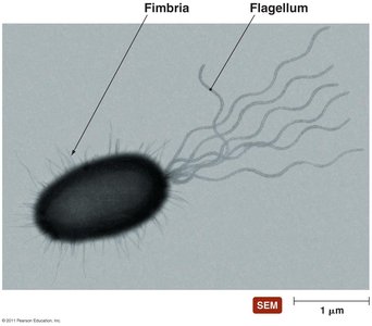
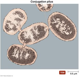
Pili
Pili are longer than fimbriae but shorter than flagella. They are typically present in small numbers and mediate the transfer of DNA between cells (conjugation).
Composed of pilin
Also known as conjugation pili
Function: DNA transfer (conjugation)
Bacterial Cell Walls
Structure and Function
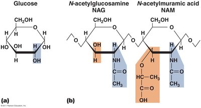
Bacterial cell walls provide structural support, protect against osmotic forces, and contribute to cell shape. They are composed primarily of peptidoglycan.
Peptidoglycan: Polymer of N-acetylglucosamine (NAG) and N-acetylmuramic acid (NAM)
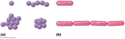
Shapes: Cocci (spherical), bacilli (rod-shaped), etc.
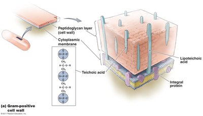
Gram-Positive Cell Walls
Gram-positive bacteria have thick peptidoglycan layers and contain teichoic acids. They stain purple in the Gram stain procedure.
Thick peptidoglycan layer
Teichoic acids: Unique polyalcohols
Mycolic acid: In acid-fast bacteria, aids in desiccation resistance

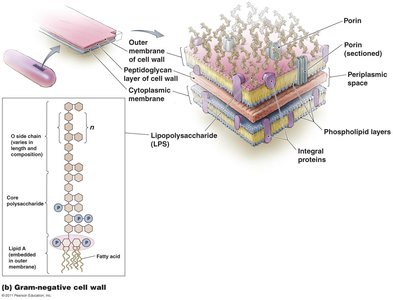
Gram-Negative Cell Walls
Gram-negative bacteria have a thin peptidoglycan layer and an outer membrane containing lipopolysaccharide (LPS). They stain pink in the Gram stain procedure.
Thin peptidoglycan layer
Outer membrane: Contains LPS, phospholipids, proteins
Impediment to treatment: LPS can be toxic and hinder antibiotic entry

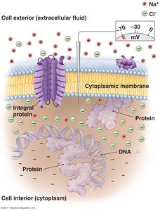
Bacterial Cytoplasmic Membranes
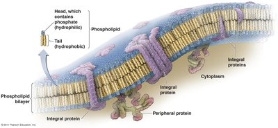
Structure
The cytoplasmic membrane is a phospholipid bilayer with embedded proteins, described by the fluid mosaic model.
Phospholipid bilayer: Hydrophilic heads, hydrophobic tails
Integral and peripheral proteins: Various functions
Function
The membrane is selectively permeable, involved in energy storage, and maintains concentration and electrical gradients.
Energy storage
Harvest light energy (photosynthetic bacteria)
Selectively permeable
Proteins allow substance transport
Maintain gradients
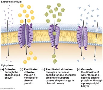
Passive Processes
Substances move across membranes via passive processes such as diffusion, facilitated diffusion, and osmosis.
Diffusion: Movement of molecules from high to low concentration
Facilitated diffusion: Uses channel proteins
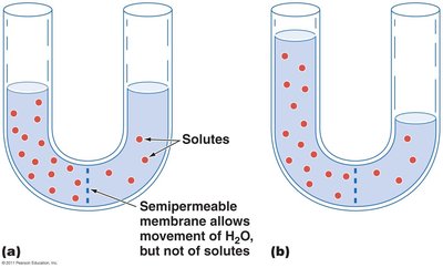
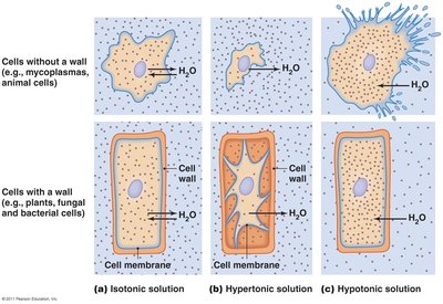
Osmosis: Movement of water across a semipermeable membrane
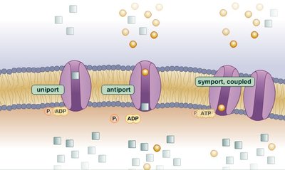
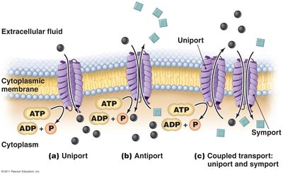
Active Processes
Active transport requires energy (ATP) to move substances against concentration gradients. Group translocation chemically modifies substances during transport.
Active transport: Uniport, antiport, symport mechanisms
Group translocation: Substance is chemically modified during transport
Cytoplasm of Bacteria
Cytosol, Inclusions, and Endospores
The cytoplasm contains cytosol (liquid), inclusions (reserve deposits), and endospores (defensive structures).
Cytosol: Liquid portion
Inclusions: Storage of chemicals
Endospores: Survival structures in harsh conditions
Nonmembranous Organelles
Bacterial cells contain ribosomes (sites of protein synthesis) and a cytoskeleton (maintains cell shape).
Ribosomes: 70S, composed of protein and RNA
Cytoskeleton: Simple, helical structure
Archaeal Cell Structure
External Structures
Archaea possess glycocalyces, flagella, fimbriae, and hami, with unique features compared to bacteria.
Glycocalyces: Biofilm formation, adherence
Flagella: Basal body, hook, filament; differences from bacterial flagella
Fimbriae and hami: Attachment structures
Cell Walls and Cytoplasmic Membranes
Most archaea have cell walls without peptidoglycan, composed of specialized polysaccharides and proteins. All have cytoplasmic membranes.
Cell wall: No peptidoglycan
Cytoplasmic membrane: Maintains gradients, controls import/export
Cytoplasm of Archaea
Archaeal cytoplasm is similar to bacteria but differs in ribosomal proteins, metabolic enzymes, and genetic code.
70S ribosomes
Fibrous cytoskeleton
Circular DNA
Differences: Ribosomal proteins, metabolic enzymes, genetic code
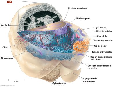
Eukaryotic Cell Structure
External Structures
Eukaryotic cells have glycocalyces that are less organized than prokaryotic capsules. They aid in cell anchoring, surface strengthening, dehydration protection, and cell recognition.
Cell Walls and Cytoplasmic Membranes
Cell walls are present in fungi, algae, plants, and some protozoa, composed of various polysaccharides. All eukaryotic cells have a fluid mosaic cytoplasmic membrane with steroid lipids and membrane rafts.
Cellulose: Plant cell walls
Chitin, glucomannan: Fungal cell walls
Various polysaccharides: Algal cell walls
Membrane rafts: Regions of lipids and proteins
Endocytosis
Eukaryotic cells can internalize substances via endocytosis, a process involving membrane invagination.
Flagella and Cilia
Eukaryotic flagella differ structurally and functionally from prokaryotic flagella. Cilia are shorter and more numerous, used for movement and moving substances past the cell surface.
Flagella: Tubulin microtubules, undulate rhythmically
Cilia: Coordinated beating, propels cells
Other Nonmembranous Organelles
Eukaryotic cells contain ribosomes (80S), cytoskeleton (tubulin, actin, intermediate filaments), centrioles, and centrosomes.
Ribosomes: 80S (60S + 40S subunits)
Cytoskeleton: Extensive network
Centrioles: Role in mitosis, cytokinesis, flagella/cilia formation
Centrosome: Region containing centrioles
Membranous Organelles
Eukaryotic cells possess several membrane-bound organelles, each with specialized functions.
Nucleus: Contains DNA, nucleoplasm, chromatin, nucleoli, nuclear envelope, nuclear pores
Endoplasmic reticulum: SER (lipid synthesis), RER (protein synthesis)
Golgi body: Processes and packages molecules for export
Lysosomes: Catabolic enzymes
Peroxisomes: Degrade poisonous wastes
Vacuoles and vesicles: Storage and transport
Mitochondria: ATP production, contains 70S ribosomes and circular DNA
Chloroplasts: Light-harvesting, contains 70S ribosomes and DNA
Endosymbiotic Theory
The endosymbiotic theory proposes that eukaryotes originated from a symbiotic relationship between small aerobic prokaryotes and larger anaerobic prokaryotes, leading to the development of mitochondria and chloroplasts.
Small aerobic prokaryotes became internal parasites
Larger cell became dependent on parasites for ATP production
Aerobic prokaryotes evolved into mitochondria
Similar scenario for chloroplasts
Additional info: The endosymbiotic theory is supported by the presence of 70S ribosomes and circular DNA in mitochondria and chloroplasts, similar to prokaryotes.
